2J3S | pdb_00002j3s

Crystal structure of the human filamin A Ig domains 19 to 21


Experimental Data Snapshot

  • Method: X-RAY DIFFRACTION
  • Resolution: 2.50 Å
  • R-Value Free: 
    0.297 (Depositor), 0.285 (DCC) 
  • R-Value Work: 
    0.252 (Depositor), 0.248 (DCC) 
  • R-Value Observed: 
    0.257 (Depositor) 

Starting Model: experimental
View more details

wwPDB Validation 3D Report Full Report

Validation slider image for 2J3S

This is version 1.2 of the entry. See complete history

Literature

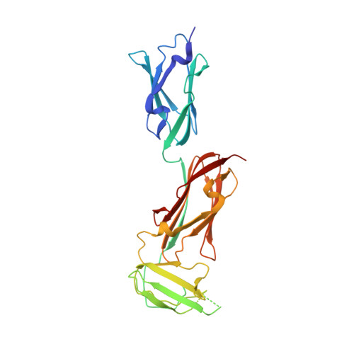
Structure of Three Tandem Filamin Domains Reveals Auto-Inhibition of Ligand-Binding.

Lad, Y.Kiema, T.-R.Jiang, P.Pentikanen, O.T.Coles, C.H.Campbell, I.D.Calderwood, D.A.Ylanne, J.

(2007) EMBO J 26: 3993

  • DOI: https://doi.org/10.1038/sj.emboj.7601827
  • Primary Citation Related Structures: 
    2J3S

  • PubMed Abstract: 

    Human filamins are large actin-crosslinking proteins composed of an N-terminal actin-binding domain followed by 24 Ig-like domains (IgFLNs), which interact with numerous transmembrane receptors and cytosolic signaling proteins. Here we report the 2.5 A resolution structure of a three-domain fragment of human filamin A (IgFLNa19-21). The structure reveals an unexpected domain arrangement, with IgFLNa20 partially unfolded bringing IgFLNa21 into close proximity to IgFLNa19. Notably the N-terminus of IgFLNa20 forms a beta-strand that associates with the CD face of IgFLNa21 and occupies the binding site for integrin adhesion receptors. Disruption of this IgFLNa20-IgFLNa21 interaction enhances filamin binding to integrin beta-tails. Structural and functional analysis of other IgFLN domains suggests that auto-inhibition by adjacent IgFLN domains may be a general mechanism controlling filamin-ligand interactions. This can explain the increased integrin binding of filamin splice variants and provides a mechanism by which ligand binding might impact filamin structure.


  • Organizational Affiliation
    • Department of Pharmacology and Interdepartmental Program in Vascular Biology and Transplantation, Yale University School of Medicine, New Haven, CT 06520, USA.

Macromolecule Content 

  • Total Structure Weight: 61.8 kDa 
  • Atom Count: 3,779 
  • Modeled Residue Count: 500 
  • Deposited Residue Count: 576 
  • Unique protein chains: 1

Macromolecules

Find similar proteins by:|  3D Structure
Entity ID: 1
MoleculeChains  Sequence LengthOrganismDetailsImage
FILAMIN-A
A, B
288Homo sapiensMutation(s): 0 
UniProt & NIH Common Fund Data Resources
Find proteins for P21333 (Homo sapiens)
Explore P21333 
Go to UniProtKB:  P21333
PHAROS:  P21333
GTEx:  ENSG00000196924 
Entity Groups
Sequence Clusters30% Identity50% Identity70% Identity90% Identity95% Identity100% Identity
UniProt GroupP21333
Sequence Annotations
Expand
Reference Sequence

Small Molecules

Ligands 3 Unique
IDChains Name / Formula / InChI Key2D Diagram3D Interactions
GOL

Query on GOL


Download:Ideal Coordinates CCD File
I [auth B]GLYCEROL
C3 H8 O3
PEDCQBHIVMGVHV-UHFFFAOYSA-N
DIO

Query on DIO


Download:Ideal Coordinates CCD File
D [auth A],
E [auth A],
G [auth B],
H [auth B]
1,4-DIETHYLENE DIOXIDE
C4 H8 O2
RYHBNJHYFVUHQT-UHFFFAOYSA-N
BR

Query on BR


Download:Ideal Coordinates CCD File
C [auth A],
F [auth B]
BROMIDE ION
Br
CPELXLSAUQHCOX-UHFFFAOYSA-M

Experimental Data & Validation

Experimental Data

  • Method: X-RAY DIFFRACTION
  • Resolution: 2.50 Å
  • R-Value Free:  0.297 (Depositor), 0.285 (DCC) 
  • R-Value Work:  0.252 (Depositor), 0.248 (DCC) 
  • R-Value Observed: 0.257 (Depositor) 
Space Group: C 2 2 21
Unit Cell:
Length ( Å )Angle ( ˚ )
a = 72.28α = 90
b = 78.39β = 90
c = 229.04γ = 90
Software Package:
Software NamePurpose
REFMACrefinement
XDSdata reduction
XDSdata scaling
PHASERphasing

Structure Validation

View Full Validation Report


Entry History 

Deposition Data

Revision History  (Full details and data files)

  • Version 1.0: 2007-10-16
    Type: Initial release
  • Version 1.1: 2011-07-13
    Changes: Advisory, Refinement description, Version format compliance
  • Version 1.2: 2023-12-13
    Changes: Data collection, Database references, Other, Refinement description