2J3P | pdb_00002j3p

crystal structure of rat FGF1 at 1.4 A


Experimental Data Snapshot

  • Method: X-RAY DIFFRACTION
  • Resolution: 1.40 Å
  • R-Value Free: 
    0.229 (Depositor), 0.225 (DCC) 
  • R-Value Work: 
    0.202 (Depositor), 0.199 (DCC) 
  • R-Value Observed: 
    0.204 (Depositor) 

Starting Model: experimental
View more details

wwPDB Validation 3D Report Full Report

Validation slider image for 2J3P

This is version 1.3 of the entry. See complete history

Literature

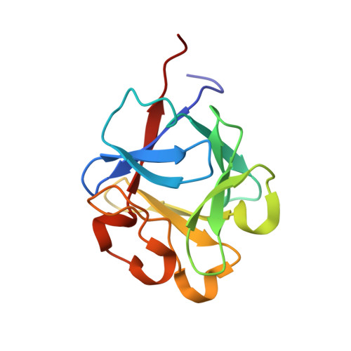
Structure of Rat Acidic Fibroblast Growth Factor at 1.4 A Resolution.

Kulahin, N.Kiselyov, V.Kochoyan, A.Kristensen, O.Kastrup, J.S.Berezin, V.Bock, E.Gajhede, M.

(2007) Acta Crystallogr Sect F Struct Biol Cryst Commun 63: 65

  • DOI: https://doi.org/10.1107/S1744309107003144
  • Primary Citation Related Structures: 
    2J3P

  • PubMed Abstract: 

    Fibroblast growth factors (FGFs) constitute a family of 22 structurally related heparin-binding polypeptides that are involved in the regulation of cell growth, survival, differentiation and migration. Here, a 1.4 A resolution X-ray structure of rat FGF1 is presented. Two molecules are present in the asymmetric unit of the crystal and they coordinate a total of five sulfate ions. The structures of human, bovine and newt FGF1 have been published previously. Human and rat FGF1 are found to have very similar structures.


  • Organizational Affiliation
    • Protein Laboratory, Institute of Molecular Pathology, Panum Institute, Blegdamsvej 3C, DK-2200 Copenhagen, Denmark. kulahin@plab.ku.dk

Macromolecule Content 

  • Total Structure Weight: 30.83 kDa 
  • Atom Count: 2,477 
  • Modeled Residue Count: 266 
  • Deposited Residue Count: 268 
  • Unique protein chains: 1

Macromolecules

Find similar proteins by:|  3D Structure
Entity ID: 1
MoleculeChains  Sequence LengthOrganismDetailsImage
HEPARIN-BINDING GROWTH FACTOR 1
A, B
134Rattus norvegicusMutation(s): 0 
UniProt
Find proteins for P61149 (Rattus norvegicus)
Explore P61149 
Go to UniProtKB:  P61149
Entity Groups
Sequence Clusters30% Identity50% Identity70% Identity90% Identity95% Identity100% Identity
UniProt GroupP61149
Sequence Annotations
Expand
Reference Sequence

Experimental Data & Validation

Experimental Data

  • Method: X-RAY DIFFRACTION
  • Resolution: 1.40 Å
  • R-Value Free:  0.229 (Depositor), 0.225 (DCC) 
  • R-Value Work:  0.202 (Depositor), 0.199 (DCC) 
  • R-Value Observed: 0.204 (Depositor) 
Space Group: P 21 21 21
Unit Cell:
Length ( Å )Angle ( ˚ )
a = 42.882α = 90
b = 61.655β = 90
c = 88.856γ = 90
Software Package:
Software NamePurpose
REFMACrefinement
MOSFLMdata reduction
SCALAdata scaling
PHASERphasing

Structure Validation

View Full Validation Report


Entry History 

Deposition Data

Revision History  (Full details and data files)

  • Version 1.0: 2007-02-13
    Type: Initial release
  • Version 1.1: 2011-05-08
    Changes: Version format compliance
  • Version 1.2: 2011-07-13
    Changes: Version format compliance
  • Version 1.3: 2023-12-13
    Changes: Data collection, Database references, Other, Refinement description