2IVJ | pdb_00002ivj

Isopenicillin N Synthase From Aspergillus Nidulans (Anaerobic Ac- cyclopropylglycine Fe Complex)


Experimental Data Snapshot

  • Method: X-RAY DIFFRACTION
  • Resolution: 1.46 Å
  • R-Value Free: 
    0.167 (Depositor), 0.179 (DCC) 
  • R-Value Work: 
    0.141 (Depositor), 0.152 (DCC) 
  • R-Value Observed: 
    0.142 (Depositor) 

wwPDB Validation 3D Report Full Report

Validation slider image for 2IVJ

Ligand Structure Quality Assessment 


This is version 1.2 of the entry. See complete history

Literature

Interactions of Isopenicillin N Synthase with Cyclopropyl-Containing Substrate Analogues Reveal New Mechanistic Insight.

Howard-Jones, A.R.Elkins, J.M.Clifton, I.J.Roach, P.L.Adlington, R.M.Baldwin, J.E.Rutledge, P.J.

(2007) Biochemistry 46: 4755

  • DOI: https://doi.org/10.1021/bi062314q
  • Primary Citation Related Structures: 
    2IVI, 2IVJ

  • PubMed Abstract: 

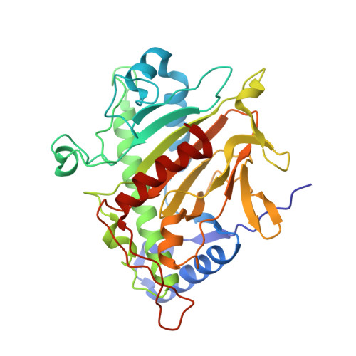
    Isopenicillin N synthase (IPNS), a non-heme iron oxidase central to penicillin and cephalosporin biosynthesis, catalyzes an energetically demanding chemical transformation to produce isopenicillin N from the tripeptide delta-(l-alpha-aminoadipoyl)-l-cysteinyl-d-valine (ACV). We describe the synthesis of two cyclopropyl-containing tripeptide analogues, delta-(l-alpha-aminoadipoyl)-l-cysteinyl-beta-methyl-d-cyclopropylglycine and delta-(l-alpha-aminoadipoyl)-l-cysteinyl-d-cyclopropylglycine, designed as probes for the mechanism of IPNS. We have solved the X-ray crystal structures of these substrates in complex with IPNS and propose a revised mechanism for the IPNS-mediated turnover of these compounds. Relative to the previously determined IPNS-Fe(II)-ACV structure, key differences exist in substrate orientation and water occupancy, which allow for an explanation of the differences in reactivity of these substrates.


  • Organizational Affiliation
    • Chemistry Research Laboratory, University of Oxford, Mansfield Road, Oxford OX1 3TA, UK.

Macromolecule Content 

  • Total Structure Weight: 38.17 kDa 
  • Atom Count: 3,131 
  • Modeled Residue Count: 329 
  • Deposited Residue Count: 331 
  • Unique protein chains: 1

Macromolecules

Find similar proteins by:|  3D Structure
Entity ID: 1
MoleculeChains  Sequence LengthOrganismDetailsImage
ISOPENICILLIN N SYNTHETASE331Aspergillus nidulans FGSC A4Mutation(s): 0 
EC: 1.21.3.1
UniProt
Find proteins for P05326 (Emericella nidulans (strain FGSC A4 / ATCC 38163 / CBS 112.46 / NRRL 194 / M139))
Explore P05326 
Go to UniProtKB:  P05326
Entity Groups
Sequence Clusters30% Identity50% Identity70% Identity90% Identity95% Identity100% Identity
UniProt GroupP05326
Sequence Annotations
Expand
Reference Sequence

Experimental Data & Validation

Experimental Data

  • Method: X-RAY DIFFRACTION
  • Resolution: 1.46 Å
  • R-Value Free:  0.167 (Depositor), 0.179 (DCC) 
  • R-Value Work:  0.141 (Depositor), 0.152 (DCC) 
  • R-Value Observed: 0.142 (Depositor) 
Space Group: P 21 21 21
Unit Cell:
Length ( Å )Angle ( ˚ )
a = 46.663α = 90
b = 71.182β = 90
c = 100.877γ = 90
Software Package:
Software NamePurpose
REFMACrefinement
MOSFLMdata reduction
SCALAdata scaling

Structure Validation

View Full Validation Report


Ligand Structure Quality Assessment 


Entry History 

Deposition Data

Revision History  (Full details and data files)

  • Version 1.0: 2007-04-10
    Type: Initial release
  • Version 1.1: 2014-02-19
    Changes: Database references, Derived calculations, Other, Version format compliance
  • Version 1.2: 2024-05-08
    Changes: Data collection, Database references, Derived calculations, Other, Source and taxonomy