2IS1 | pdb_00002is1

Crystal structure of UvrD-DNA-SO4 complex


Experimental Data Snapshot

  • Method: X-RAY DIFFRACTION
  • Resolution: 2.90 Å
  • R-Value Free: 
    0.295 (Depositor), 0.285 (DCC) 
  • R-Value Work: 
    0.236 (Depositor), 0.229 (DCC) 
  • R-Value Observed: 
    0.236 (Depositor) 

Starting Model: experimental
View more details

wwPDB Validation 3D Report Full Report

Validation slider image for 2IS1

This is version 1.5 of the entry. See complete history

Literature

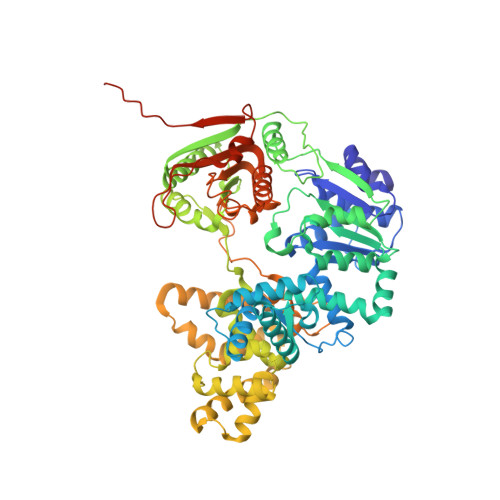
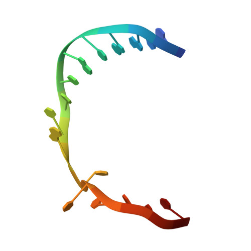
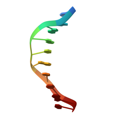
UvrD helicase unwinds DNA one base pair at a time by a two-part power stroke.

Lee, J.Y.Yang, W.

(2006) Cell 127: 1349-1360

  • DOI: https://doi.org/10.1016/j.cell.2006.10.049
  • Primary Citation Related Structures: 
    2IS1, 2IS2, 2IS4, 2IS6

  • PubMed Abstract: 

    Helicases use the energy derived from nucleoside triphosphate hydrolysis to unwind double helices in essentially every metabolic pathway involving nucleic acids. Earlier crystal structures have suggested that DNA helicases translocate along a single-stranded DNA in an inchworm fashion. We report here a series of crystal structures of the UvrD helicase complexed with DNA and ATP hydrolysis intermediates. These structures reveal that ATP binding alone leads to unwinding of 1 base pair by directional rotation and translation of the DNA duplex, and ADP and Pi release leads to translocation of the developing single strand. Thus DNA unwinding is achieved by a two-part power stroke in a combined wrench-and-inchworm mechanism. The rotational angle and translational distance of DNA define the unwinding step to be 1 base pair per ATP hydrolyzed. Finally, a gateway for ssDNA translocation and an alternative strand-displacement mode may explain the varying step sizes reported previously.


  • Organizational Affiliation
    • Laboratory of Molecular Biology, National Institute of Diabetes and Digestive and Kidney Diseases, National Institutes of Health, Bethesda, MD 20892, USA.

Macromolecule Content 

  • Total Structure Weight: 173.51 kDa 
  • Atom Count: 10,896 
  • Modeled Residue Count: 1,324 
  • Deposited Residue Count: 1,414 
  • Unique protein chains: 1
  • Unique nucleic acid chains: 2

Macromolecules


Find similar proteins by:|  3D Structure
Entity ID: 3
MoleculeChains  Sequence LengthOrganismDetailsImage
DNA helicase IIE [auth A],
F [auth B]
680Escherichia coliMutation(s): 1 
Gene Names: uvrD
EC: 3.6.1 (PDB Primary Data), 5.6.2.4 (UniProt)
UniProt
Find proteins for P03018 (Escherichia coli (strain K12))
Explore P03018 
Go to UniProtKB:  P03018
Entity Groups
Sequence Clusters30% Identity50% Identity70% Identity90% Identity95% Identity100% Identity
UniProt GroupP03018
Sequence Annotations
Expand
Reference Sequence
Find similar nucleic acids by:  Sequence
Entity ID: 1
MoleculeChains LengthOrganismImage
5'-D(*GP*CP*AP*GP*TP*GP*CP*TP*CP*GP*TP*TP*TP*TP*TP*TP*T)-3'A [auth C],
C [auth E]
17N/A
Sequence Annotations
Expand
Reference Sequence
Find similar nucleic acids by:  Sequence
Entity ID: 2
MoleculeChains LengthOrganismImage
5'-D(*CP*GP*AP*GP*CP*AP*CP*TP*GP*C)-3'B [auth D],
D [auth F]
10N/A
Sequence Annotations
Expand
Reference Sequence

Small Molecules

Ligands 3 Unique
IDChains Name / Formula / InChI Key2D Diagram3D Interactions
HG

Query on HG


Download:Ideal Coordinates CCD File
G [auth A],
H [auth A],
I [auth A],
M [auth B],
N [auth B]
MERCURY (II) ION
Hg
BQPIGGFYSBELGY-UHFFFAOYSA-N
SO4

Query on SO4


Download:Ideal Coordinates CCD File
J [auth A],
K [auth A],
O [auth B]
SULFATE ION
O4 S
QAOWNCQODCNURD-UHFFFAOYSA-L
GOL

Query on GOL


Download:Ideal Coordinates CCD File
L [auth A],
P [auth B]
GLYCEROL
C3 H8 O3
PEDCQBHIVMGVHV-UHFFFAOYSA-N

Experimental Data & Validation

Experimental Data

  • Method: X-RAY DIFFRACTION
  • Resolution: 2.90 Å
  • R-Value Free:  0.295 (Depositor), 0.285 (DCC) 
  • R-Value Work:  0.236 (Depositor), 0.229 (DCC) 
  • R-Value Observed: 0.236 (Depositor) 
Space Group: P 21 21 2
Unit Cell:
Length ( Å )Angle ( ˚ )
a = 152.451α = 90
b = 94.49β = 90
c = 136.443γ = 90
Software Package:
Software NamePurpose
CNSrefinement
MAR345data collection
HKL-2000data reduction
HKL-2000data scaling
AMoREphasing

Structure Validation

View Full Validation Report


Entry History 

Deposition Data

Revision History  (Full details and data files)

  • Version 1.0: 2007-01-09
    Type: Initial release
  • Version 1.1: 2008-05-01
    Changes: Version format compliance
  • Version 1.2: 2011-07-13
    Changes: Non-polymer description, Version format compliance
  • Version 1.3: 2017-10-18
    Changes: Refinement description
  • Version 1.4: 2021-10-20
    Changes: Database references, Derived calculations
  • Version 1.5: 2023-08-30
    Changes: Data collection, Refinement description