2IMW | pdb_00002imw

Mechanism of Template-Independent Nucleotide Incorporation Catalyzed by a Template-Dependent DNA Polymerase


Experimental Data Snapshot

  • Method: X-RAY DIFFRACTION
  • Resolution: 2.05 Å
  • R-Value Free: 
    0.284 (Depositor), 0.272 (DCC) 
  • R-Value Work: 
    0.230 (Depositor), 0.219 (DCC) 
  • R-Value Observed: 
    0.232 (Depositor) 

Starting Model: experimental
View more details

wwPDB Validation 3D Report Full Report

Validation slider image for 2IMW

Ligand Structure Quality Assessment 


This is version 1.7 of the entry. See complete history

Literature

Mechanism of Template-independent Nucleotide Incorporation Catalyzed by a Template-dependent DNA Polymerase.

Fiala, K.A.Brown, J.A.Ling, H.Kshetry, A.K.Zhang, J.Taylor, J.S.Yang, W.Suo, Z.

(2007) J Mol Biology 365: 590-602

  • DOI: https://doi.org/10.1016/j.jmb.2006.10.008
  • Primary Citation Related Structures: 
    2IMW

  • PubMed Abstract: 

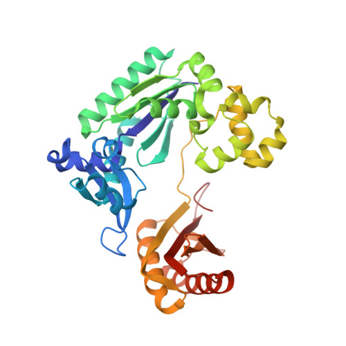
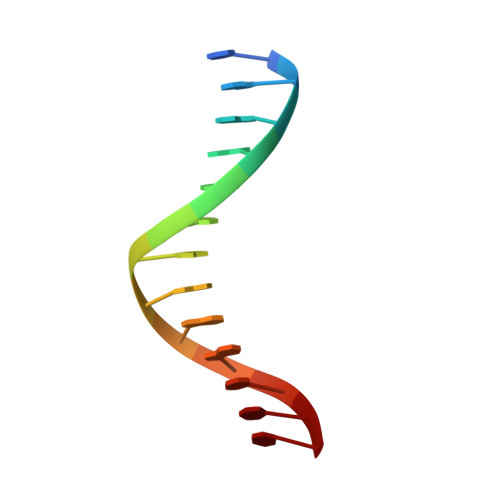
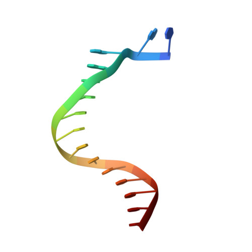
    Numerous template-dependent DNA polymerases are capable of catalyzing template-independent nucleotide additions onto blunt-end DNA. Such non-canonical activity has been hypothesized to increase the genomic hypermutability of retroviruses including human immunodeficiency viruses. Here, we employed pre-steady state kinetics and X-ray crystallography to establish a mechanism for blunt-end additions catalyzed by Sulfolobus solfataricus Dpo4. Our kinetic studies indicated that the first blunt-end dATP incorporation was 80-fold more efficient than the second, and among natural deoxynucleotides, dATP was the preferred substrate due to its stronger intrahelical base-stacking ability. Such base-stacking contributions are supported by the 41-fold higher ground-state binding affinity of a nucleotide analog, pyrene nucleoside 5'-triphosphate, which lacks hydrogen bonding ability but possesses four conjugated aromatic rings. A 2.05 A resolution structure of Dpo4*(blunt-end DNA)*ddATP revealed that the base and sugar of the incoming ddATP, respectively, stack against the 5'-base of the opposite strand and the 3'-base of the elongating strand. This unprecedented base-stacking pattern can be applied to subsequent blunt-end additions only if all incorporated dAMPs are extrahelical, leading to predominantly single non-templated dATP incorporation.


  • Organizational Affiliation
    • Department of Biochemistry, The Ohio State University, Columbus, OH 43210, USA.

Macromolecule Content 

  • Total Structure Weight: 49.24 kDa 
  • Atom Count: 3,690 
  • Modeled Residue Count: 376 
  • Deposited Residue Count: 376 
  • Unique protein chains: 1
  • Unique nucleic acid chains: 2

Macromolecules


Find similar proteins by:|  3D Structure
Entity ID: 3
MoleculeChains  Sequence LengthOrganismDetailsImage
DNA polymerase IVC [auth P]348Saccharolobus solfataricusMutation(s): 0 
Gene Names: dbin
EC: 2.7.7.7
UniProt
Find proteins for Q97W02 (Saccharolobus solfataricus (strain ATCC 35092 / DSM 1617 / JCM 11322 / P2))
Explore Q97W02 
Go to UniProtKB:  Q97W02
Entity Groups
Sequence Clusters30% Identity50% Identity70% Identity90% Identity95% Identity100% Identity
UniProt GroupQ97W02
Sequence Annotations
Expand
Reference Sequence
Find similar nucleic acids by:  Sequence
Entity ID: 1
MoleculeChains LengthOrganismImage
5'-D(*GP*GP*GP*GP*GP*AP*AP*GP*GP*AP*TP*TP*C)-3'A [auth S]13N/A
Sequence Annotations
Expand
Reference Sequence
Find similar nucleic acids by:  Sequence
Entity ID: 2
MoleculeChains LengthOrganismImage
5'-D(*TP*AP*GP*AP*AP*TP*CP*CP*TP*TP*CP*CP*CP*CP*C)-3'B [auth T]15N/A
Sequence Annotations
Expand
Reference Sequence

Small Molecules

Ligands 3 Unique
IDChains Name / Formula / InChI Key2D Diagram3D Interactions
DDS

Query on DDS


Download:Ideal Coordinates CCD File
F [auth P]2',3'-dideoxyadenosine triphosphate
C10 H16 N5 O11 P3
OAKPWEUQDVLTCN-NKWVEPMBSA-N
EDO

Query on EDO


Download:Ideal Coordinates CCD File
G [auth P]
H [auth P]
I [auth P]
J [auth P]
K [auth P]
G [auth P],
H [auth P],
I [auth P],
J [auth P],
K [auth P],
L [auth P]
1,2-ETHANEDIOL
C2 H6 O2
LYCAIKOWRPUZTN-UHFFFAOYSA-N
CA

Query on CA


Download:Ideal Coordinates CCD File
D [auth P],
E [auth P]
CALCIUM ION
Ca
BHPQYMZQTOCNFJ-UHFFFAOYSA-N

Experimental Data & Validation

Experimental Data

  • Method: X-RAY DIFFRACTION
  • Resolution: 2.05 Å
  • R-Value Free:  0.284 (Depositor), 0.272 (DCC) 
  • R-Value Work:  0.230 (Depositor), 0.219 (DCC) 
  • R-Value Observed: 0.232 (Depositor) 
Space Group: P 21 21 2
Unit Cell:
Length ( Å )Angle ( ˚ )
a = 98.057α = 90
b = 101.857β = 90
c = 52.38γ = 90
Software Package:
Software NamePurpose
REFMACrefinement
SCALEPACKdata scaling
CNSrefinement
DENZOdata reduction
PDB_EXTRACTdata extraction
HKL-2000data collection
CNSphasing

Structure Validation

View Full Validation Report


Ligand Structure Quality Assessment 


Entry History 

Deposition Data

Revision History  (Full details and data files)

  • Version 1.0: 2007-01-16
    Type: Initial release
  • Version 1.1: 2008-05-01
    Changes: Version format compliance
  • Version 1.2: 2011-07-13
    Changes: Advisory, Version format compliance
  • Version 1.3: 2017-10-18
    Changes: Refinement description
  • Version 1.4: 2018-04-04
    Changes: Data collection
  • Version 1.5: 2019-07-24
    Changes: Data collection, Derived calculations, Refinement description
  • Version 1.6: 2023-08-30
    Changes: Data collection, Database references, Derived calculations, Refinement description
  • Version 1.7: 2024-12-25
    Changes: Advisory, Derived calculations, Structure summary