2ILZ | pdb_00002ilz

Crystal structure of poliovirus polymerase complexed with GTP and Mn2+


Experimental Data Snapshot

  • Method: X-RAY DIFFRACTION
  • Resolution: 2.50 Å
  • R-Value Free: 
    0.255 (Depositor), 0.246 (DCC) 
  • R-Value Work: 
    0.230 (Depositor), 0.222 (DCC) 

Starting Model: experimental
View more details

wwPDB Validation 3D Report Full Report

Validation slider image for 2ILZ

Ligand Structure Quality Assessment 


This is version 1.5 of the entry. See complete history

Literature

Stabilization of Poliovirus Polymerase by NTP Binding and Fingers-Thumb Interactions.

Thompson, A.A.Albertini, R.A.Peersen, O.B.

(2007) J Mol Biology 366: 1459-1474

  • DOI: https://doi.org/10.1016/j.jmb.2006.11.070
  • Primary Citation Related Structures: 
    2ILY, 2ILZ, 2IM0, 2IM1, 2IM2, 2IM3

  • PubMed Abstract: 

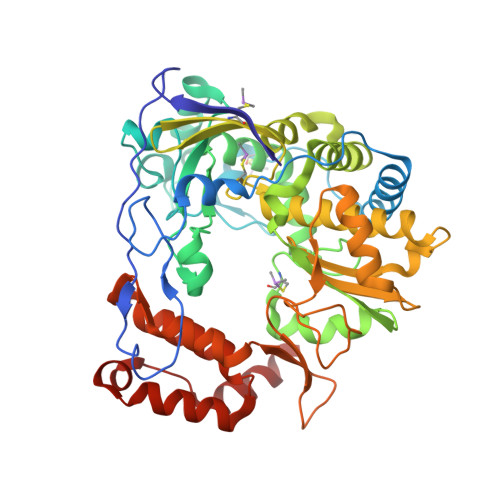
    The viral RNA-dependent RNA polymerases show a conserved structure where the fingers domain interacts with the top of the thumb domain to create a tunnel through which nucleotide triphosphates reach the active site. We have solved the crystal structures of poliovirus polymerase (3D(pol)) in complex with all four NTPs, showing that they all bind in a common pre-insertion site where the phosphate groups are not yet positioned over the active site. The NTPs interact with both the fingers and palm domains, forming bridging interactions that explain the increased thermal stability of 3D(pol) in the presence of NTPs. We have also examined the importance of the fingers-thumb domain interaction for the function and structural stability of 3D(pol). Results from thermal denaturation experiments using circular dichroism and 2-anilino-6-napthaline-sulfonate (ANS) fluorescence show that 3D(pol) has a melting temperature of only approximately 40 degrees C. NTP binding stabilizes the protein and increases the melting by 5-6 degrees C while mutations in the fingers-thumb domain interface destabilize the protein and reduce the melting point by as much as 6 degrees C. In particular, the burial of Phe30 and Phe34 from the tip of the index finger into a pocket at the top of the thumb and the presence of Trp403 on the thumb domain are key interactions required to maintain the structural integrity of the polymerase. The data suggest the fingers domain has significant conformational flexibility and exists in a highly dynamic molten globule state at physiological temperature. The role of the enclosed active site motif as a structural scaffold for constraining the fingers domain and accommodating conformational changes in 3D(pol) and other viral polymerases during the catalytic cycle is discussed.


  • Organizational Affiliation
    • Program in Cellular and Molecular Biology, Colorado State University, Fort Collins, CO 80523-1870, USA.

Macromolecule Content 

  • Total Structure Weight: 53.97 kDa 
  • Atom Count: 3,946 
  • Modeled Residue Count: 461 
  • Deposited Residue Count: 461 
  • Unique protein chains: 1

Macromolecules

Find similar proteins by:|  3D Structure
Entity ID: 1
MoleculeChains  Sequence LengthOrganismDetailsImage
poliovirus polymerase461Human poliovirus 1 MahoneyMutation(s): 6 
Gene Names: 3D
EC: 2.7.7.48
UniProt
Find proteins for P03300 (Poliovirus type 1 (strain Mahoney))
Explore P03300 
Go to UniProtKB:  P03300
Entity Groups
Sequence Clusters30% Identity50% Identity70% Identity90% Identity95% Identity100% Identity
UniProt GroupP03300
Sequence Annotations
Expand
Reference Sequence

Small Molecules

Ligands 4 Unique
IDChains Name / Formula / InChI Key2D Diagram3D Interactions
GTP

Query on GTP


Download:Ideal Coordinates CCD File
F [auth A]GUANOSINE-5'-TRIPHOSPHATE
C10 H16 N5 O14 P3
XKMLYUALXHKNFT-UUOKFMHZSA-N
ACY

Query on ACY


Download:Ideal Coordinates CCD File
G [auth A],
H [auth A],
I [auth A],
J [auth A],
K [auth A]
ACETIC ACID
C2 H4 O2
QTBSBXVTEAMEQO-UHFFFAOYSA-N
MN

Query on MN


Download:Ideal Coordinates CCD File
B [auth A],
C [auth A]
MANGANESE (II) ION
Mn
WAEMQWOKJMHJLA-UHFFFAOYSA-N
NA

Query on NA


Download:Ideal Coordinates CCD File
D [auth A],
E [auth A]
SODIUM ION
Na
FKNQFGJONOIPTF-UHFFFAOYSA-N
Modified Residues  1 Unique
IDChains TypeFormula2D DiagramParent
CAS
Query on CAS
A
L-PEPTIDE LINKINGC5 H12 As N O2 SCYS

Experimental Data & Validation

Experimental Data

  • Method: X-RAY DIFFRACTION
  • Resolution: 2.50 Å
  • R-Value Free:  0.255 (Depositor), 0.246 (DCC) 
  • R-Value Work:  0.230 (Depositor), 0.222 (DCC) 
Space Group: P 65
Unit Cell:
Length ( Å )Angle ( ˚ )
a = 128.159α = 90
b = 128.159β = 90
c = 112.61γ = 120
Software Package:
Software NamePurpose
CrystalCleardata collection
CNSrefinement
d*TREKdata reduction
d*TREKdata scaling
CNSphasing

Structure Validation

View Full Validation Report


Ligand Structure Quality Assessment 


Entry History 

Deposition Data

Revision History  (Full details and data files)

  • Version 1.0: 2006-12-12
    Type: Initial release
  • Version 1.1: 2008-05-01
    Changes: Version format compliance
  • Version 1.2: 2011-07-13
    Changes: Source and taxonomy, Version format compliance
  • Version 1.3: 2021-10-20
    Changes: Database references, Derived calculations
  • Version 1.4: 2023-08-30
    Changes: Data collection, Refinement description
  • Version 1.5: 2024-10-30
    Changes: Structure summary