2ILL

Anomalous substructure of Titin-A168169


Experimental Data Snapshot

  • Method: X-RAY DIFFRACTION
  • Resolution: 2.20 Å
  • R-Value Free: 0.243 
  • R-Value Work: 0.212 
  • R-Value Observed: 0.212 

wwPDB Validation   3D Report Full Report


This is version 1.3 of the entry. See complete history


Literature

On the routine use of soft X-rays in macromolecular crystallography. Part IV. Efficient determination of anomalous substructures in biomacromolecules using longer X-ray wavelengths

Mueller-Dieckmann, C.Panjikar, S.Schmidt, A.Mueller, S.Kuper, J.Geerlof, A.Wilmanns, M.Singh, R.K.Tucker, P.A.Weiss, M.S.

(2007) Acta Crystallogr D Biol Crystallogr 63: 366-380

  • DOI: https://doi.org/10.1107/S0907444906055624
  • Primary Citation of Related Structures:  
    2G4H, 2G4I, 2G4J, 2G4K, 2G4L, 2G4M, 2G4N, 2G4O, 2G4P, 2G4Q, 2G4R, 2G4S, 2G4T, 2G4U, 2G4V, 2G4W, 2G4X, 2G4Y, 2G4Z, 2G51, 2G52, 2G55, 2ILL

  • PubMed Abstract: 

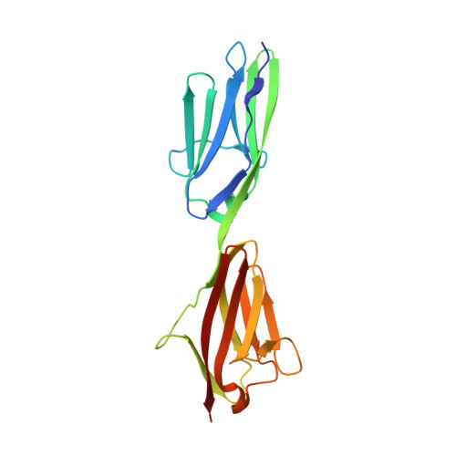
    23 different crystal forms of 19 different biological macromolecules were examined with respect to their anomalously scattering substructures using diffraction data collected at a wavelength of 2.0 A (6.2 keV). In more than 90% of the cases the substructure was found to contain more than just the protein S atoms. The data presented suggest that chloride, sulfate, phosphate or metal ions from the buffer or even from the purification protocol are frequently bound to the protein molecule and that these ions are often overlooked, especially if they are not bound at full occupancy. Thus, in order to fully describe the macromolecule under study, it seems desirable that any structure determination be complemented with a long-wavelength data set.


  • Organizational Affiliation

    EMBL Hamburg Outstation, c/o DESY, Notkestrasse 85, D-22603 Hamburg, Germany.


Macromolecules
Find similar proteins by:  (by identity cutoff)  |  3D Structure
Entity ID: 1
MoleculeChains Sequence LengthOrganismDetailsImage
Titin195Homo sapiensMutation(s): 0 
EC: 2.7.11.1
UniProt & NIH Common Fund Data Resources
Find proteins for Q8WZ42 (Homo sapiens)
Explore Q8WZ42 
Go to UniProtKB:  Q8WZ42
PHAROS:  Q8WZ42
GTEx:  ENSG00000155657 
Entity Groups  
Sequence Clusters30% Identity50% Identity70% Identity90% Identity95% Identity100% Identity
UniProt GroupQ8WZ42
Sequence Annotations
Expand
  • Reference Sequence
Experimental Data & Validation

Experimental Data

  • Method: X-RAY DIFFRACTION
  • Resolution: 2.20 Å
  • R-Value Free: 0.243 
  • R-Value Work: 0.212 
  • R-Value Observed: 0.212 
  • Space Group: I 2 2 2
Unit Cell:
Length ( Å )Angle ( ˚ )
a = 69.26α = 90
b = 89.12β = 90
c = 103.55γ = 90
Software Package:
Software NamePurpose
REFMACrefinement
HKL-2000data collection
HKL-2000data reduction
CCP4data scaling

Structure Validation

View Full Validation Report


Entry History 

Deposition Data

Revision History  (Full details and data files)

  • Version 1.0: 2007-02-20
    Type: Initial release
  • Version 1.1: 2008-05-01
    Changes: Version format compliance
  • Version 1.2: 2011-07-13
    Changes: Advisory, Version format compliance
  • Version 1.3: 2024-03-13
    Changes: Data collection, Database references, Derived calculations