2IHU | pdb_00002ihu

Carboxyethylarginine synthase from Streptomyces clavuligerus: putative reaction intermediate complex


Experimental Data Snapshot

  • Method: X-RAY DIFFRACTION
  • Resolution: 2.05 Å
  • R-Value Free: 
    0.201 (Depositor), 0.203 (DCC) 
  • R-Value Work: 
    0.171 (Depositor), 0.175 (DCC) 
  • R-Value Observed: 
    0.172 (Depositor) 

Starting Model: experimental
View more details

wwPDB Validation 3D Report Full Report

Validation slider image for 2IHU

This is version 1.2 of the entry. See complete history

Literature

Structural and mechanistic studies on N(2)-(2-carboxyethyl)arginine synthase.

Caines, M.E.Sorensen, J.L.Schofield, C.J.

(2009) Biochem Biophys Res Commun 385: 512-517

  • DOI: https://doi.org/10.1016/j.bbrc.2009.05.095
  • Primary Citation Related Structures: 
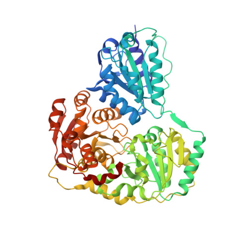
    2IHT, 2IHU, 2IHV

  • PubMed Abstract: 

    N(2)-(2-Carboxyethyl)arginine synthase (CEAS), an unusual thiamin diphosphate (ThDP)-dependent enzyme, catalyses the committed step in the biosynthesis of the b-lactamase inhibitor clavulanic acid in Streptomyces clavuligerus. Crystal structures of tetrameric CEAS-ThDP in complex with the substrate analogues 5-guanidinovaleric acid (GVA) and tartrate, and a structure reflecting a possible enol(ate)-ThDP reaction intermediate are described. The structures suggest overlapping binding sites for the substrates D-glyceraldehyde-3-phosphate (D-G3P) and L-arginine, and are consistent with the proposed CEAS mechanism in which D-G3P binds at the active site and reacts to form an alpha,beta-unsaturated intermediate,which subsequently undergoes (1,4)-Michael addition with the alpha-amino group of L-arginine. Additional solution studies are presented which probe the amino acid substrate tolerance of CEAS, providing further insight into the L-arginine binding site. These findings may facilitate the engineering of CEAS towards the synthesis of alternative beta-amino acid products.


  • Organizational Affiliation
    • Department of Chemistry, University of Oxford, Chemistry Research Laboratory, Oxford OX13TA, United Kingdom.

Macromolecule Content 

  • Total Structure Weight: 246.81 kDa 
  • Atom Count: 18,287 
  • Modeled Residue Count: 2,216 
  • Deposited Residue Count: 2,292 
  • Unique protein chains: 1

Macromolecules

Find similar proteins by:|  3D Structure
Entity ID: 1
MoleculeChains  Sequence LengthOrganismDetailsImage
Carboxyethylarginine synthase
A, B, C, D
573Streptomyces clavuligerusMutation(s): 0 
Gene Names: ceas2
EC: 2.5.1.66
UniProt
Find proteins for Q9LCV9 (Streptomyces clavuligerus)
Explore Q9LCV9 
Go to UniProtKB:  Q9LCV9
Entity Groups
Sequence Clusters30% Identity50% Identity70% Identity90% Identity95% Identity100% Identity
UniProt GroupQ9LCV9
Sequence Annotations
Expand
Reference Sequence

Small Molecules

Ligands 6 Unique
IDChains Name / Formula / InChI Key2D Diagram3D Interactions
TP8

Query on TP8


Download:Ideal Coordinates CCD File
Q [auth C]5-(2-{[HYDROXY(PHOSPHONOOXY)PHOSPHORYL]OXY}ETHYL)-2-[(1Z)-1-HYDROXY-3-(PHOSPHONOOXY)PROP-1-EN-1-YL]-3-{[(4Z)-4-IMINO-2- METHYL-4,5-DIHYDROPYRIMIDIN-5-YL]METHYL}-4-METHYL-1,3-THIAZOL-3-IUM
C15 H21 N4 O12 P3 S
IYJJFPCOWJABBY-AOCMNYQTSA-L
TP9

Query on TP9


Download:Ideal Coordinates CCD File
H [auth A],
L [auth B],
R [auth C],
U [auth D]
(3Z)-4-{[(4-AMINO-2-METHYLPYRIMIDIN-5-YL)METHYL]AMINO}-3-MERCAPTOPENT-3-EN-1-YL TRIHYDROGEN DIPHOSPHATE
C11 H18 N4 O7 P2 S
WTQDUFKDQLXDPH-YFHOEESVSA-L
TAR

Query on TAR


Download:Ideal Coordinates CCD File
I [auth A],
V [auth D]
D(-)-TARTARIC ACID
C4 H6 O6
FEWJPZIEWOKRBE-LWMBPPNESA-N
GOL

Query on GOL


Download:Ideal Coordinates CCD File
M [auth B]GLYCEROL
C3 H8 O3
PEDCQBHIVMGVHV-UHFFFAOYSA-N
K

Query on K


Download:Ideal Coordinates CCD File
F [auth A]
G [auth A]
K [auth B]
O [auth C]
P [auth C]
F [auth A],
G [auth A],
K [auth B],
O [auth C],
P [auth C],
T [auth D]
POTASSIUM ION
K
NPYPAHLBTDXSSS-UHFFFAOYSA-N
MG

Query on MG


Download:Ideal Coordinates CCD File
E [auth A],
J [auth B],
N [auth C],
S [auth D]
MAGNESIUM ION
Mg
JLVVSXFLKOJNIY-UHFFFAOYSA-N

Experimental Data & Validation

Experimental Data

  • Method: X-RAY DIFFRACTION
  • Resolution: 2.05 Å
  • R-Value Free:  0.201 (Depositor), 0.203 (DCC) 
  • R-Value Work:  0.171 (Depositor), 0.175 (DCC) 
  • R-Value Observed: 0.172 (Depositor) 
Space Group: P 21 21 21
Unit Cell:
Length ( Å )Angle ( ˚ )
a = 119.691α = 90
b = 127.863β = 90
c = 197.15γ = 90
Software Package:
Software NamePurpose
SCALAdata scaling
REFMACrefinement
PDB_EXTRACTdata extraction
MOSFLMdata reduction
REFMACphasing

Structure Validation

View Full Validation Report


Entry History 

Deposition Data

Revision History  (Full details and data files)

  • Version 1.0: 2007-11-27
    Type: Initial release
  • Version 1.1: 2011-07-13
    Changes: Non-polymer description, Version format compliance
  • Version 1.2: 2023-08-30
    Changes: Data collection, Database references, Derived calculations, Refinement description, Structure summary