2I21 | pdb_00002i21

Bacteriorhodopsin/lipid complex, T46V mutant


Experimental Data Snapshot

  • Method: X-RAY DIFFRACTION
  • Resolution: 1.84 Å
  • R-Value Free: 
    0.260 (Depositor) 
  • R-Value Work: 
    0.195 (Depositor), 0.180 (DCC) 
  • R-Value Observed: 
    0.197 (Depositor) 

Starting Model: experimental
View more details

wwPDB Validation   3D Report Full Report


Ligand Structure Quality Assessment 

Created with Raphaël 2.3.0Worse 01 BetterLigand structure goodness of fit to experimental dataBest fitted LI1Click on this verticalbar to view detailsBest fitted SQUClick on this verticalbar to view detailsBest fitted RETClick on this verticalbar to view details

This is version 1.5 of the entry. See complete history


Literature

Propagating Structural Perturbation Inside Bacteriorhodopsin: Crystal Structures of the M State and the D96A and T46V Mutants.

Lanyi, J.K.Schobert, B.

(2006) Biochemistry 45: 12003-12010

  • DOI: https://doi.org/10.1021/bi061310i
  • Primary Citation of Related Structures:  
    2I1X, 2I20, 2I21

  • PubMed Abstract: 

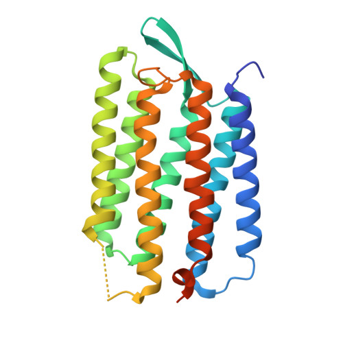
    The X-ray diffraction structure of the non-illuminated D96A bacteriorhodopsin mutant reveals structural changes as far away as 15 A from residue 96, at the retinal, Trp-182, Ala-215, and waters 501, 402, and 401. The Asp-to-Ala side-chain replacement breaks its hydrogen bond with Thr-46, and the resulting separation of the cytoplasmic ends of helices B and C is communicated to the retinal region through a chain of covalent and hydrogen bonds. The unexpected long-range consequences of the D96A mutation include breaking the hydrogen bond between O of Ala-215 and water 501 and the formation of a new hydrogen bond between water molecules 401 and 402 in the extracellular region. Because in the T46V mutant a new water molecule appears at Asp-96 and its hydrogen-bond to Ile-45 replaces Thr-46 as its link to helix B, the separation of helices B and C is smaller than that in D96A, and there are no atomic displacements elsewhere in the protein. Propagation of conformational changes along the chain between the retinal and Thr-46 had been observed earlier in the crystal structures of the D96N and E204Q mutants but in the trapped M state. Consistent with the perturbation of the retinal region in D96A, little change of the Thr-46 region occurs between the non-illuminated and M states of this mutant. It appears that a local perturbation can propagate along a track in both directions between the retinal and the Asp-96/Thr-46 pair, either from photoisomerization of the retinal in the wild-type protein in one case or from the D96A mutation in the other.


  • Organizational Affiliation

    Department of Physiology and Biophysics, University of California, Irvine, California 92697, USA. jlanyi@orion.oac.uci.edu


Macromolecules
Find similar proteins by:  (by identity cutoff)  |  3D Structure
Entity ID: 1
MoleculeChains Sequence LengthOrganismDetailsImage
Bacteriorhodopsin249Halobacterium salinarumMutation(s): 1 
Membrane Entity: Yes 
UniProt
Find proteins for P02945 (Halobacterium salinarum (strain ATCC 700922 / JCM 11081 / NRC-1))
Explore P02945 
Go to UniProtKB:  P02945
Entity Groups  
Sequence Clusters30% Identity50% Identity70% Identity90% Identity95% Identity100% Identity
UniProt GroupP02945
Sequence Annotations
Expand
  • Reference Sequence
Small Molecules
Ligands 3 Unique
IDChains Name / Formula / InChI Key2D Diagram3D Interactions
LI1
Query on LI1

Download Ideal Coordinates CCD File 
C [auth A]
D [auth A]
E [auth A]
F [auth A]
G [auth A]
1-[2,6,10.14-TETRAMETHYL-HEXADECAN-16-YL]-2-[2,10,14-TRIMETHYLHEXADECAN-16-YL]GLYCEROL
C42 H86 O3
YERVUJAKCNBGCR-BIHSMRAKSA-N
SQU
Query on SQU

Download Ideal Coordinates CCD File 
P [auth A]2,10,23-TRIMETHYL-TETRACOSANE
C27 H56
ZADHKSJXSZBQFB-HHHXNRCGSA-N
RET
Query on RET

Download Ideal Coordinates CCD File 
B [auth A]RETINAL
C20 H28 O
NCYCYZXNIZJOKI-OVSJKPMPSA-N
Experimental Data & Validation

Experimental Data

  • Method: X-RAY DIFFRACTION
  • Resolution: 1.84 Å
  • R-Value Free:  0.260 (Depositor) 
  • R-Value Work:  0.195 (Depositor), 0.180 (DCC) 
  • R-Value Observed: 0.197 (Depositor) 
Space Group: P 63
Unit Cell:
Length ( Å )Angle ( ˚ )
a = 61.069α = 90
b = 61.069β = 90
c = 110.534γ = 120
Software Package:
Software NamePurpose
SHELXmodel building
SHELXL-97refinement
ADSCdata collection
HKL-2000data reduction
HKL-2000data scaling
SHELXphasing

Structure Validation

View Full Validation Report


Ligand Structure Quality Assessment 

Created with Raphaël 2.3.0Worse 01 BetterLigand structure goodness of fit to experimental dataBest fitted LI1Click on this verticalbar to view detailsBest fitted SQUClick on this verticalbar to view detailsBest fitted RETClick on this verticalbar to view details

Entry History 

Deposition Data

Revision History  (Full details and data files)

  • Version 1.0: 2006-10-10
    Type: Initial release
  • Version 1.1: 2008-05-01
    Changes: Version format compliance
  • Version 1.2: 2011-07-13
    Changes: Derived calculations, Version format compliance
  • Version 1.3: 2021-10-20
    Changes: Database references, Derived calculations
  • Version 1.4: 2023-08-30
    Changes: Data collection, Refinement description
  • Version 1.5: 2024-10-09
    Changes: Structure summary