2I13 | pdb_00002i13

Aart, a six finger zinc finger designed to recognize ANN triplets


Experimental Data Snapshot

  • Method: X-RAY DIFFRACTION
  • Resolution: 1.96 Å
  • R-Value Free: 
    0.249 (Depositor), 0.246 (DCC) 
  • R-Value Work: 
    0.203 (Depositor), 0.210 (DCC) 
  • R-Value Observed: 
    0.203 (Depositor) 

wwPDB Validation 3D Report Full Report

Validation slider image for 2I13

This is version 1.3 of the entry. See complete history

Literature

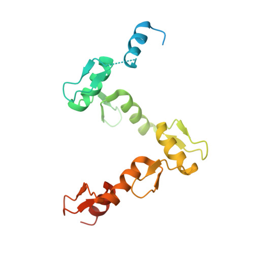
Structure of Aart, a Designed Six-finger Zinc Finger Peptide, Bound to DNA.

Segal, D.J.Crotty, J.W.Bhakta, M.S.Barbas, C.F.Horton, N.C.

(2006) J Mol Biology 363: 405-421

  • DOI: https://doi.org/10.1016/j.jmb.2006.08.016
  • Primary Citation Related Structures: 
    2I13

  • PubMed Abstract: 

    Cys2-His2 zinc fingers are one of the most common types of DNA-binding domains. Modifications to zinc-finger binding specificity have recently enabled custom DNA-binding proteins to be designed to a wide array of target sequences. We present here a 1.96 A structure of Aart, a designed six-zinc finger protein, bound to a consensus DNA target site. This is the first structure of a designed protein with six fingers, and was intended to provide insights into the unusual affinity and specificity characteristics of this protein. Most protein-DNA contacts were found to be consistent with expectations, while others were unanticipated or insufficient to explain specificity. Several were unexpectedly mediated by glycerol, water molecules or amino acid-base stacking interactions. These results challenge some conventional concepts of recognition, particularly the finding that triplets containing 5'A, C, or T are typically not specified by direct interaction with the amino acid in position 6 of the recognition helix.


  • Organizational Affiliation
    • UC Davis Genome Center and Department of Pharmacology, University of California, Davis, CA 95616, USA. djsegal@ucdavis.edu

Macromolecule Content 

  • Total Structure Weight: 71.08 kDa 
  • Atom Count: 4,534 
  • Modeled Residue Count: 383 
  • Deposited Residue Count: 468 
  • Unique protein chains: 1
  • Unique nucleic acid chains: 2

Macromolecules


Find similar proteins by:|  3D Structure
Entity ID: 3
MoleculeChains  Sequence LengthOrganismDetailsImage
AartE [auth A],
F [auth B]
190Mus musculusMutation(s): 0 
UniProt & NIH Common Fund Data Resources
Find proteins for Q07230 (Mus musculus)
Explore Q07230 
Go to UniProtKB:  Q07230
IMPC:  MGI:99176
Entity Groups
Sequence Clusters30% Identity50% Identity70% Identity90% Identity95% Identity100% Identity
UniProt GroupQ07230
Sequence Annotations
Expand
Reference Sequence
Find similar nucleic acids by:  Sequence
Entity ID: 1
MoleculeChains LengthOrganismImage
5'-D(*CP*AP*GP*AP*TP*GP*TP*AP*GP*GP*GP*AP*AP*AP*AP*GP*CP*CP*CP*GP*GP*G)-3'A [auth C],
C [auth E]
22N/A
Sequence Annotations
Expand
Reference Sequence
Find similar nucleic acids by:  Sequence
Entity ID: 2
MoleculeChains LengthOrganismImage
5'-D(*GP*CP*CP*CP*GP*GP*GP*CP*TP*TP*TP*TP*CP*CP*CP*TP*AP*CP*AP*TP*CP*T)-3'B [auth D],
D [auth F]
22N/A
Sequence Annotations
Expand
Reference Sequence

Small Molecules

Ligands 2 Unique
IDChains Name / Formula / InChI Key2D Diagram3D Interactions
GOL

Query on GOL


Download:Ideal Coordinates CCD File
G [auth C],
H [auth C],
I [auth E],
J [auth E]
GLYCEROL
C3 H8 O3
PEDCQBHIVMGVHV-UHFFFAOYSA-N
ZN

Query on ZN


Download:Ideal Coordinates CCD File
K [auth A]
L [auth A]
M [auth A]
N [auth A]
O [auth A]
K [auth A],
L [auth A],
M [auth A],
N [auth A],
O [auth A],
P [auth A],
Q [auth B],
R [auth B],
S [auth B],
T [auth B],
U [auth B]
ZINC ION
Zn
PTFCDOFLOPIGGS-UHFFFAOYSA-N

Experimental Data & Validation

Experimental Data

  • Method: X-RAY DIFFRACTION
  • Resolution: 1.96 Å
  • R-Value Free:  0.249 (Depositor), 0.246 (DCC) 
  • R-Value Work:  0.203 (Depositor), 0.210 (DCC) 
  • R-Value Observed: 0.203 (Depositor) 
Space Group: P 1
Unit Cell:
Length ( Å )Angle ( ˚ )
a = 41.586α = 75.53
b = 72.054β = 83.8
c = 74.212γ = 72.67
Software Package:
Software NamePurpose
REFMACrefinement
PDB_EXTRACTdata extraction
HKL-2000data reduction
HKL-2000data scaling
SOLVEphasing

Structure Validation

View Full Validation Report


Entry History 

Deposition Data

Revision History  (Full details and data files)

  • Version 1.0: 2006-10-03
    Type: Initial release
  • Version 1.1: 2008-05-01
    Changes: Version format compliance
  • Version 1.2: 2011-07-13
    Changes: Non-polymer description, Version format compliance
  • Version 1.3: 2024-02-21
    Changes: Advisory, Data collection, Database references, Derived calculations