


Crystal structure of possible sugar phosphatase, HAD superfamily (ZP_00311070.1) from CYTOPHAGA HUTCHINSONII ATCC 33406 at 2.10 A resolution
Joint Center for Structural Genomics (JCSG)To be published.
Experimental Data Snapshot
wwPDB Validation 3D Report Full Report

Entity ID: 1 | |||||
|---|---|---|---|---|---|
| Molecule | Chains | Sequence Length | Organism | Details | Image |
| Predicted sugar phosphatases of the HAD superfamily | 284 | Cytophaga hutchinsonii | Mutation(s): 9 Gene Names: ZP_00311070.1 | 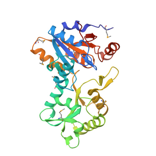 | |
| Ligands 4 Unique | |||||
|---|---|---|---|---|---|
| ID | Chains | Name / Formula / InChI Key | 2D Diagram | 3D Interactions | |
| EPE Download:Ideal Coordinates CCD File | FA [auth C], L [auth A], NA [auth D], V [auth B] | 4-(2-HYDROXYETHYL)-1-PIPERAZINE ETHANESULFONIC ACID C8 H18 N2 O4 S JKMHFZQWWAIEOD-UHFFFAOYSA-N |  | ||
| EDO Download:Ideal Coordinates CCD File | AA [auth B] GA [auth C] HA [auth C] IA [auth C] JA [auth C] | 1,2-ETHANEDIOL C2 H6 O2 LYCAIKOWRPUZTN-UHFFFAOYSA-N |  | ||
| CL Download:Ideal Coordinates CCD File | CA [auth C] DA [auth C] EA [auth C] F [auth A] G [auth A] | CHLORIDE ION Cl VEXZGXHMUGYJMC-UHFFFAOYSA-M |  | ||
| MG Download:Ideal Coordinates CCD File | BA [auth C], E [auth A], LA [auth D], P [auth B] | MAGNESIUM ION Mg JLVVSXFLKOJNIY-UHFFFAOYSA-N |  | ||
| Modified Residues 1 Unique | |||||
|---|---|---|---|---|---|
| ID | Chains | Type | Formula | 2D Diagram | Parent |
| MSE Query on MSE | A, B, C, D | L-PEPTIDE LINKING | C5 H11 N O2 Se |  | MET |
| Length ( Å ) | Angle ( ˚ ) |
|---|---|
| a = 65.624 | α = 90 |
| b = 119.462 | β = 90 |
| c = 151.274 | γ = 90 |
| Software Name | Purpose |
|---|---|
| MolProbity | model building |
| SHELX | phasing |
| REFMAC | refinement |
| SCALA | data scaling |
| PDB_EXTRACT | data extraction |
| MOSFLM | data reduction |
| CCP4 | data scaling |
| SHELXD | phasing |
| autoSHARP | phasing |


