2HVQ | pdb_00002hvq

Structure of Adenylated full-length T4 RNA Ligase 2


Experimental Data Snapshot

  • Method: X-RAY DIFFRACTION
  • Resolution: 2.40 Å
  • R-Value Free: 
    0.274 (Depositor), 0.204 (DCC) 
  • R-Value Work: 
    0.211 (Depositor), 0.214 (DCC) 

Starting Model: experimental
View more details

wwPDB Validation 3D Report Full Report

Validation slider image for 2HVQ

This is version 1.4 of the entry. See complete history

Literature

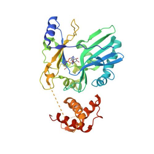
RNA Ligase Structures Reveal the Basis for RNA Specificity and Conformational Changes that Drive Ligation Forward.

Nandakumar, J.Shuman, S.Lima, C.D.

(2006) Cell 127: 71-84

  • DOI: https://doi.org/10.1016/j.cell.2006.08.038
  • Primary Citation Related Structures: 
    2HVQ, 2HVR, 2HVS

  • PubMed Abstract: 

    T4 RNA ligase 2 (Rnl2) and kinetoplastid RNA editing ligases exemplify a family of RNA repair enzymes that seal 3'OH/5'PO(4) nicks in duplex RNAs via ligase adenylylation (step 1), AMP transfer to the nick 5'PO(4) (step 2), and attack by the nick 3'OH on the 5'-adenylylated strand to form a phosphodiester (step 3). Crystal structures are reported for Rnl2 at discrete steps along this pathway: the covalent Rnl2-AMP intermediate; Rnl2 bound to an adenylylated nicked duplex, captured immediately following step 2; and Rnl2 at an adenylylated nick in a state poised for step 3. These structures illuminate the stereochemistry of nucleotidyl transfer and reveal how remodeling of active-site contacts and conformational changes propel the ligation reaction forward. Mutational analysis and comparison of nick-bound structures of Rnl2 and human DNA ligase I highlight common and divergent themes of substrate recognition that can explain their specialization for RNA versus DNA repair.


  • Organizational Affiliation
    • Structural Biology Program, Sloan-Kettering Institute, New York, NY 10021, USA.

Macromolecule Content 

  • Total Structure Weight: 38.07 kDa 
  • Atom Count: 2,637 
  • Modeled Residue Count: 317 
  • Deposited Residue Count: 335 
  • Unique protein chains: 1

Macromolecules

Find similar proteins by:|  3D Structure
Entity ID: 1
MoleculeChains  Sequence LengthOrganismDetailsImage
Hypothetical 37.6 kDa protein in Gp24-hoc intergenic region335Tequatrovirus T4Mutation(s): 1 
Gene Names: Y10A24.1
EC: 6.5.1.3
UniProt
Find proteins for P32277 (Enterobacteria phage T4)
Explore P32277 
Go to UniProtKB:  P32277
Entity Groups
Sequence Clusters30% Identity50% Identity70% Identity90% Identity95% Identity100% Identity
UniProt GroupP32277
Sequence Annotations
Expand
Reference Sequence

Small Molecules

Modified Residues  1 Unique
IDChains TypeFormula2D DiagramParent
APK
Query on APK
A
L-PEPTIDE LINKINGC16 H26 N7 O7 PLYS

Experimental Data & Validation

Experimental Data

  • Method: X-RAY DIFFRACTION
  • Resolution: 2.40 Å
  • R-Value Free:  0.274 (Depositor), 0.204 (DCC) 
  • R-Value Work:  0.211 (Depositor), 0.214 (DCC) 
Space Group: P 21 21 21
Unit Cell:
Length ( Å )Angle ( ˚ )
a = 48.02α = 90
b = 57.85β = 90
c = 119.49γ = 90
Software Package:
Software NamePurpose
DENZOdata reduction
SCALEPACKdata scaling
CNSrefinement
PDB_EXTRACTdata extraction
MOLREPphasing

Structure Validation

View Full Validation Report


Entry History 

Deposition Data

Revision History  (Full details and data files)

  • Version 1.0: 2006-10-17
    Type: Initial release
  • Version 1.1: 2008-05-01
    Changes: Version format compliance
  • Version 1.2: 2011-07-13
    Changes: Version format compliance
  • Version 1.3: 2017-10-18
    Changes: Refinement description
  • Version 1.4: 2023-08-30
    Changes: Data collection, Database references, Derived calculations, Refinement description