Solution structure of polymerase mu's BRCT Domain reveals an element essential for its role in nonhomologous end joining.
DeRose, E.F., Clarkson, M.W., Gilmore, S.A., Galban, C.J., Tripathy, A., Havener, J.M., Mueller, G.A., Ramsden, D.A., London, R.E., Lee, A.L.(2007) Biochemistry 46: 12100-12110
- PubMed: 17915942 Search on PubMedSearch on PubMed Central
- DOI: https://doi.org/10.1021/bi7007728
- Primary Citation Related Structures:
2HTF - PubMed Abstract:
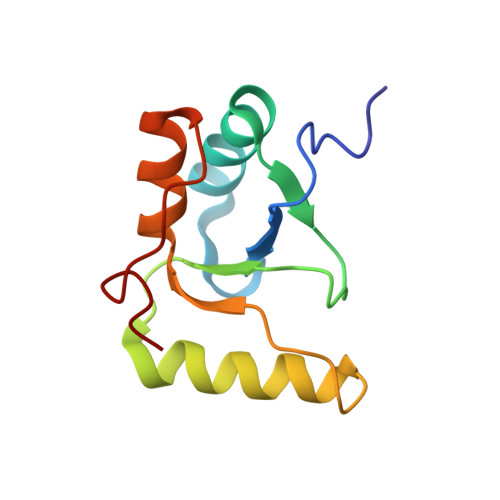
The solution structure and dynamics of the BRCT domain from human DNA polymerase mu, implicated in repair of chromosome breaks by nonhomologous end joining (NHEJ), has been determined using NMR methods. BRCT domains are typically involved in protein-protein interactions between factors required for the cellular response to DNA damage. The pol mu BRCT domain is atypical in that, unlike other reported BRCT structures, the pol mu BRCT is neither part of a tandem grouping, nor does it appear to form stable homodimers. Although the sequence of the pol mu BRCT domain has some unique characteristics, particularly the presence of >10% proline residues, it forms the characteristic alphabetaalpha sandwich, in which three alpha helices are arrayed around a central four-stranded beta-sheet. The structure of helix alpha1 is characterized by two solvent-exposed hydrophobic residues, F46 and L50, suggesting that this element may play a role in mediating interactions of pol mu with other proteins. Consistent with this argument, mutation of these residues, as well as the proximal, conserved residue R43, specifically blocked the ability of pol mu to efficiently work together with NHEJ factors Ku and XRCC4-ligase IV to join noncomplementary ends together in vitro. The structural, dynamic, and biochemical evidence reported here identifies a functional surface in the pol mu BRCT domain critical for promoting assembly and activity of the NHEJ machinery. Further, the similarity between the interaction regions of the BRCT domains of pol mu and TdT support the conclusion that they participate in NHEJ as alternate polymerases.
- Laboratory of Structural Biology, National Institute of Environmental Health Sciences, 111 T. W. Alexander Drive, Research Triangle Park, North Carolina 27709, USA.
Organizational Affiliation: