2HPS | pdb_00002hps

Crystal structure of coelenterazine-binding protein from Renilla Muelleri


Experimental Data Snapshot

  • Method: X-RAY DIFFRACTION
  • Resolution: 1.72 Å
  • R-Value Free: 
    0.229 (Depositor) 
  • R-Value Work: 
    0.199 (Depositor), 0.200 (DCC) 
  • R-Value Observed: 
    0.200 (Depositor) 

wwPDB Validation 3D Report Full Report

Validation slider image for 2HPS

Ligand Structure Quality Assessment 


This is version 1.3 of the entry. See complete history

Literature

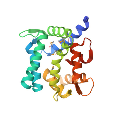
Crystal structure of coelenterazine-binding protein from Renilla muelleri at 1.7 A: why it is not a calcium-regulated photoprotein.

Stepanyuk, G.A.Liu, Z.J.Markova, S.S.Frank, L.A.Lee, J.Vysotski, E.S.Wang, B.C.

(2008) Photochem Photobiol Sci 7: 442-447

  • DOI: https://doi.org/10.1039/b716535h
  • Primary Citation Related Structures: 
    2HPS, 2HQ8

  • PubMed Abstract: 

    Bioluminescence in the sea pansy Renilla involves two distinct proteins, a Ca2+-triggered coelenterazine-binding protein (CBP), and Renilla luciferase. CBP contains one tightly bound coelenterazine molecule, which becomes available for reaction with luciferase and O2 only subsequent to Ca2+ binding. CBP belongs to the EF-hand superfamily of Ca2+-binding proteins and contains three "EF-hand" Ca2+-binding sites. The overall spatial structure of recombinant selenomethionine-labeled CBP determined at 1.7 A, is found to approximate the protein scaffold characteristic of the class of Ca2+-regulated photoproteins. Photoproteins however, catalyze molecular oxygen addition to coelenterazine producing a 2-hydroperoxycoelenterazine intermediate, which is stabilized within the binding cavity in the absence of Ca2+. Addition of Ca2+ triggers the bioluminescence reaction. However in CBP this first step of oxygen addition is not allowed. The different amino acid environments and hydrogen bond interactions within the binding cavity, are proposed to account for the different properties of the two classes of proteins.


  • Organizational Affiliation
    • Department of Biochemistry and Molecular Biology, University of Georgia, Athens, GA 30602, USA.

Macromolecule Content 

  • Total Structure Weight: 21.76 kDa 
  • Atom Count: 1,675 
  • Modeled Residue Count: 184 
  • Deposited Residue Count: 186 
  • Unique protein chains: 1

Macromolecules

Find similar proteins by:|  3D Structure
Entity ID: 1
MoleculeChains  Sequence LengthOrganismDetailsImage
coelenterazine-binding protein with bound coelenterazine186Renilla muelleriMutation(s): 0 
UniProt
Find proteins for C9V488 (Renilla muelleri)
Explore C9V488 
Go to UniProtKB:  C9V488
Entity Groups
Sequence Clusters30% Identity50% Identity70% Identity90% Identity95% Identity100% Identity
UniProt GroupC9V488
Sequence Annotations
Expand
Reference Sequence

Small Molecules

Experimental Data & Validation

Experimental Data

  • Method: X-RAY DIFFRACTION
  • Resolution: 1.72 Å
  • R-Value Free:  0.229 (Depositor) 
  • R-Value Work:  0.199 (Depositor), 0.200 (DCC) 
  • R-Value Observed: 0.200 (Depositor) 
Space Group: P 65 2 2
Unit Cell:
Length ( Å )Angle ( ˚ )
a = 55.948α = 90
b = 55.948β = 90
c = 292.202γ = 120
Software Package:
Software NamePurpose
REFMACrefinement
HKL-2000data reduction
HKL-2000data scaling
SCA2STRUCTUREphasing

Structure Validation

View Full Validation Report


Ligand Structure Quality Assessment 


Entry History 

Revision History  (Full details and data files)

  • Version 1.0: 2007-01-09
    Type: Initial release
  • Version 1.1: 2008-05-01
    Changes: Version format compliance
  • Version 1.2: 2011-07-13
    Changes: Non-polymer description, Version format compliance
  • Version 1.3: 2024-10-30
    Changes: Data collection, Database references, Derived calculations, Structure summary