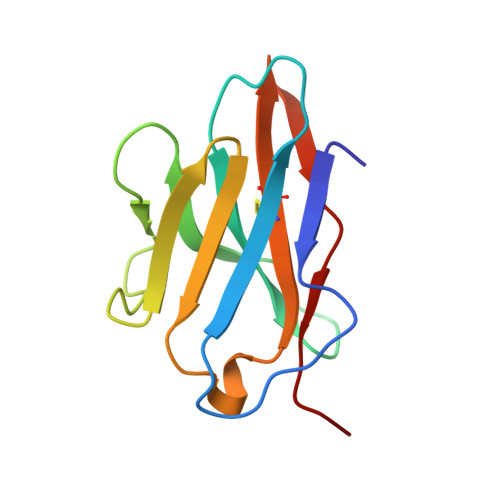
2HP4

Computational design and crystal structure of an enhanced affinity mutant human CD8-alpha-alpha co-receptor


Experimental Data Snapshot

  • Method: X-RAY DIFFRACTION
  • Resolution: 2.10 Å
  • R-Value Free: 0.233 
  • R-Value Work: 0.182 
  • R-Value Observed: 0.185 

Starting Model: experimental
View more details

wwPDB Validation   3D Report Full Report


This is version 1.4 of the entry. See complete history


Literature

Computational design and crystal structure of an enhanced affinity mutant human CD8 alphaalpha coreceptor

Cole, D.K.Rizkallah, P.J.Boulter, J.M.Sami, M.Vuidepot, A.-L.Glick, M.Gao, F.Bell, J.I.Jakobsen, B.K.Gao, G.F.

(2007) Proteins 67: 65-74

  • DOI: https://doi.org/10.1002/prot.21176
  • Primary Citation of Related Structures:  
    2HP4

  • PubMed Abstract: 

    Human CD8 is a T cell coreceptor, which binds to pHLA I and plays a pivotal role in the activation of cytotoxic T lymphocytes. Soluble recombinant CD8 alphaalpha has been shown to antagonize T cell activation, both in vitro and in vivo. However, because of a very low affinity for pHLA I, high concentrations of soluble CD8 alphaalpha are required for efficient inhibition. Based upon our knowledge of the wild-type CD8/pHLA I structure, we have designed and produced a mutated form of soluble CD8 alphaalpha that binds to pHLA I with approximately fourfold higher affinity. We have characterized the binding of the high affinity CD8 mutant using surface plasmon resonance and determined its structure at 2.1 A resolution using X-ray crystallography. The analysis of this structure suggests that the higher affinity is achieved by providing a larger side chain that allows for an optimal contact to be made between the HLA alpha3 loop and the mutated CDR-like loops of CD8.


  • Organizational Affiliation

    Nuffield Department of Clinical Medicine, John Radcliffe Hospital, Oxford University, Oxford, United Kingdom.


Macromolecules
Find similar proteins by:  (by identity cutoff)  |  3D Structure
Entity ID: 1
MoleculeChains Sequence LengthOrganismDetailsImage
T-cell surface glycoprotein CD8 alpha chain
A, B
114Homo sapiensMutation(s): 2 
UniProt & NIH Common Fund Data Resources
Find proteins for P01732 (Homo sapiens)
Explore P01732 
Go to UniProtKB:  P01732
PHAROS:  P01732
GTEx:  ENSG00000153563 
Entity Groups  
Sequence Clusters30% Identity50% Identity70% Identity90% Identity95% Identity100% Identity
UniProt GroupP01732
Sequence Annotations
Expand
  • Reference Sequence
Small Molecules
Ligands 2 Unique
IDChains Name / Formula / InChI Key2D Diagram3D Interactions
SO4
Query on SO4

Download Ideal Coordinates CCD File 
C [auth A],
D [auth A],
E [auth A],
F [auth A],
I [auth B]
SULFATE ION
O4 S
QAOWNCQODCNURD-UHFFFAOYSA-L
GOL
Query on GOL

Download Ideal Coordinates CCD File 
G [auth A],
H [auth A],
J [auth B],
K [auth B]
GLYCEROL
C3 H8 O3
PEDCQBHIVMGVHV-UHFFFAOYSA-N
Experimental Data & Validation

Experimental Data

  • Method: X-RAY DIFFRACTION
  • Resolution: 2.10 Å
  • R-Value Free: 0.233 
  • R-Value Work: 0.182 
  • R-Value Observed: 0.185 
  • Space Group: P 64
Unit Cell:
Length ( Å )Angle ( ˚ )
a = 99.723α = 90
b = 99.723β = 90
c = 55.806γ = 120
Software Package:
Software NamePurpose
SCALAdata scaling
MOLREPphasing
REFMACrefinement
PDB_EXTRACTdata extraction
MOSFLMdata reduction
CCP4data scaling

Structure Validation

View Full Validation Report


Entry History 

Deposition Data

Revision History  (Full details and data files)

  • Version 1.0: 2007-02-06
    Type: Initial release
  • Version 1.1: 2008-05-01
    Changes: Version format compliance
  • Version 1.2: 2011-07-13
    Changes: Advisory, Version format compliance
  • Version 1.3: 2021-11-10
    Changes: Database references, Derived calculations
  • Version 1.4: 2023-10-25
    Changes: Data collection, Refinement description