2HLN | pdb_00002hln

L-asparaginase from Erwinia carotovora in complex with glutamic acid


Experimental Data Snapshot

  • Method: X-RAY DIFFRACTION
  • Resolution: 2.20 Å
  • R-Value Free: 
    0.267 (Depositor), 0.262 (DCC) 
  • R-Value Work: 
    0.220 (Depositor), 0.217 (DCC) 
  • R-Value Observed: 
    0.222 (Depositor) 

Starting Model: experimental
View more details

wwPDB Validation 3D Report Full Report

Validation slider image for 2HLN

This is version 1.4 of the entry. See complete history

Literature

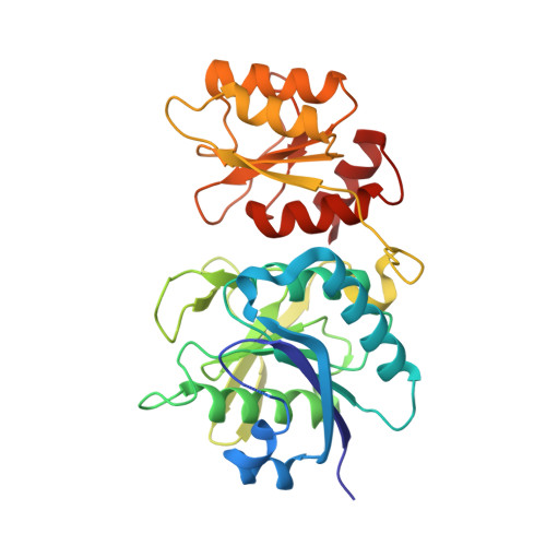
Three-dimensional structures of L-asparaginase from Erwinia carotovora complexed with aspartate and glutamate.

Kravchenko, O.V.Kislitsin, Y.A.Popov, A.N.Nikonov, S.V.Kuranova, I.P.

(2008) Acta Crystallogr D Biol Crystallogr 64: 248-256

  • DOI: https://doi.org/10.1107/S0907444907065766
  • Primary Citation Related Structures: 
    2GVN, 2HLN

  • PubMed Abstract: 

    The crystal structures of Erwinia carotovora L-asparaginase complexed with L-aspartate and L-glutamate were determined at 1.9 and 2.2 A, respectively, using the molecular-replacement method and were refined to R factors of about 21% in both cases. The positions of the ligands in the active site were located. A comparison of the new structures with the known structures of Escherichia coli L-asparaginase and Er. chrysanthemi L-asparaginase was performed. It was found that the arrangement of the ligands practically coincides in all three enzymes. The peculiarities of the quaternary structure of the enzyme, the possible role of water molecules in the enzyme action and the conformational changes during the catalyzed reaction are discussed.


  • Organizational Affiliation
    • Institute of Protein Research, Russian Academy of Sciences, 142290 Pushchino, Moscow Region, Russia.

Macromolecule Content 

  • Total Structure Weight: 416.95 kDa 
  • Atom Count: 29,749 
  • Modeled Residue Count: 3,696 
  • Deposited Residue Count: 3,924 
  • Unique protein chains: 1

Macromolecules

Find similar proteins by:|  3D Structure
Entity ID: 1
MoleculeChains  Sequence LengthOrganismDetailsImage
L-asparaginase327Pectobacterium atrosepticumMutation(s): 0 
Gene Names: lanS
EC: 3.5.1.1
UniProt
Find proteins for Q7WWK9 (Pectobacterium atrosepticum)
Explore Q7WWK9 
Go to UniProtKB:  Q7WWK9
Entity Groups
Sequence Clusters30% Identity50% Identity70% Identity90% Identity95% Identity100% Identity
UniProt GroupQ7WWK9
Sequence Annotations
Expand
Reference Sequence

Small Molecules

Ligands 2 Unique
IDChains Name / Formula / InChI Key2D Diagram3D Interactions
GLU

Query on GLU


Download:Ideal Coordinates CCD File
AA [auth K]
FA [auth L]
M [auth A]
O [auth B]
Q [auth E]
AA [auth K],
FA [auth L],
M [auth A],
O [auth B],
Q [auth E],
R [auth F],
S [auth C],
U [auth D],
V [auth G],
X [auth H],
Y [auth I],
Z [auth J]
GLUTAMIC ACID
C5 H9 N O4
WHUUTDBJXJRKMK-VKHMYHEASA-N
PEG

Query on PEG


Download:Ideal Coordinates CCD File
BA [auth K]
CA [auth K]
DA [auth K]
EA [auth K]
N [auth A]
BA [auth K],
CA [auth K],
DA [auth K],
EA [auth K],
N [auth A],
P [auth B],
T [auth C],
W [auth G]
DI(HYDROXYETHYL)ETHER
C4 H10 O3
MTHSVFCYNBDYFN-UHFFFAOYSA-N

Experimental Data & Validation

Experimental Data

  • Method: X-RAY DIFFRACTION
  • Resolution: 2.20 Å
  • R-Value Free:  0.267 (Depositor), 0.262 (DCC) 
  • R-Value Work:  0.220 (Depositor), 0.217 (DCC) 
  • R-Value Observed: 0.222 (Depositor) 
Space Group: P 1 21 1
Unit Cell:
Length ( Å )Angle ( ˚ )
a = 81.107α = 90
b = 123.675β = 90.92
c = 197.454γ = 90
Software Package:
Software NamePurpose
REFMACrefinement
PHASERphasing

Structure Validation

View Full Validation Report


Entry History 

Deposition Data

Revision History  (Full details and data files)

  • Version 1.0: 2007-07-17
    Type: Initial release
  • Version 1.1: 2008-05-01
    Changes: Version format compliance
  • Version 1.2: 2011-07-13
    Changes: Version format compliance
  • Version 1.3: 2016-02-24
    Changes: Database references
  • Version 1.4: 2023-08-30
    Changes: Data collection, Database references, Derived calculations, Refinement description