2H9S

Crystal Structure of Homo-DNA and Nature's Choice of Pentose over Hexose in the Genetic System

  • Classification: DNA
  • Mutation(s): No 

  • Deposited: 2006-06-11 Released: 2006-12-12 
  • Deposition Author(s): Egli, M., Pallan, P.S.

Experimental Data Snapshot

  • Method: X-RAY DIFFRACTION
  • Resolution: 1.75 Å
  • R-Value Free: 0.280 
  • R-Value Work: 0.240 
  • R-Value Observed: 0.240 

wwPDB Validation   3D Report Full Report


This is version 1.4 of the entry. See complete history


Literature

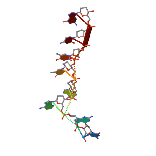
Crystal structure of homo-DNA and nature's choice of pentose over hexose in the genetic system.

Egli, M.Pallan, P.S.Pattanayek, R.Wilds, C.J.Lubini, P.Minasov, G.Dobler, M.Leumann, C.J.Eschenmoser, A.

(2006) J Am Chem Soc 128: 10847-10856

  • DOI: https://doi.org/10.1021/ja062548x
  • Primary Citation of Related Structures:  
    2H9S

  • PubMed Abstract: 

    An experimental rationalization of the structure type encountered in DNA and RNA by systematically investigating the chemical and physical properties of alternative nucleic acids has identified systems with a variety of sugar-phosphate backbones that are capable of Watson-Crick base pairing and in some cases cross-pairing with the natural nucleic acids. The earliest among the model systems tested to date, (4' --> 6')-linked oligo(2',3'-dideoxy-beta-d-glucopyranosyl)nucleotides or homo-DNA, shows stable self-pairing, but the pairing rules for the four natural bases are not the same as those in DNA. However, a complete interpretation and understanding of the properties of the hexapyranosyl (4' --> 6') family of nucleic acids has been impeded until now by the lack of detailed 3D-structural data. We have determined the crystal structure of a homo-DNA octamer. It reveals a weakly twisted right-handed duplex with a strong inclination between the hexose-phosphate backbones and base-pair axes, and highly irregular values for helical rise and twist at individual base steps. The structure allows a rationalization of the inability of allo-, altro-, and glucopyranosyl-based oligonucleotides to form stable pairing systems.


  • Organizational Affiliation

    Department of Biochemistry, School of Medicine, Vanderbilt University, Nashville, Tennessee 37232, USA. martin.egli@vanderbilt.edu


Macromolecules

Find similar nucleic acids by:  Sequence   |   3D Structure  

Entity ID: 1
MoleculeChains LengthOrganismImage
5'-D(*(XCT)P*(XGU)P*(XAD)P*(XAD)P*(XTH)P*(XTH)P*(XCT)P*(XGU))-3'
A, B
8N/A
Sequence Annotations
Expand
  • Reference Sequence
Small Molecules
Ligands 1 Unique
IDChains Name / Formula / InChI Key2D Diagram3D Interactions
MG
Query on MG

Download Ideal Coordinates CCD File 
C [auth A]MAGNESIUM ION
Mg
JLVVSXFLKOJNIY-UHFFFAOYSA-N
Experimental Data & Validation

Experimental Data

  • Method: X-RAY DIFFRACTION
  • Resolution: 1.75 Å
  • R-Value Free: 0.280 
  • R-Value Work: 0.240 
  • R-Value Observed: 0.240 
  • Space Group: P 61 2 2
Unit Cell:
Length ( Å )Angle ( ˚ )
a = 38.94α = 90
b = 38.94β = 90
c = 133.85γ = 120
Software Package:
Software NamePurpose
REFMACrefinement
HKL-2000data reduction
CNSrefinement
DENZOdata reduction
HKL-2000data scaling
CNSphasing

Structure Validation

View Full Validation Report


Entry History 

Deposition Data

Revision History  (Full details and data files)

  • Version 1.0: 2006-12-12
    Type: Initial release
  • Version 1.1: 2008-05-01
    Changes: Version format compliance
  • Version 1.2: 2011-07-13
    Changes: Version format compliance
  • Version 1.3: 2019-07-24
    Changes: Advisory, Data collection, Derived calculations, Refinement description
  • Version 1.4: 2024-02-14
    Changes: Advisory, Data collection, Database references, Derived calculations