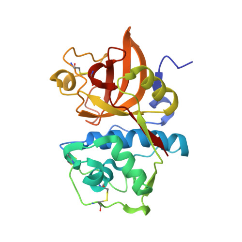
2H7J

Crystal Structure of Cathepsin S in complex with a Nonpeptidic Inhibitor.


Experimental Data Snapshot

  • Method: X-RAY DIFFRACTION
  • Resolution: 1.50 Å
  • R-Value Free: 0.180 
  • R-Value Work: 0.161 
  • R-Value Observed: 0.162 

Starting Model: experimental
View more details

wwPDB Validation   3D Report Full Report


Ligand Structure Quality Assessment 


This is version 1.3 of the entry. See complete history


Literature

Identification of selective, nonpeptidic nitrile inhibitors of cathepsin s using the substrate activity screening method.

Patterson, A.W.Wood, W.J.Hornsby, M.Lesley, S.Spraggon, G.Ellman, J.A.

(2006) J Med Chem 49: 6298-6307

  • DOI: https://doi.org/10.1021/jm060701s
  • Primary Citation of Related Structures:  
    2H7J, 2HXZ

  • PubMed Abstract: 

    The substrate activity screening method, a substrate-based fragment identification and optimization method for the development of enzyme inhibitors, was previously applied to cathepsin S to obtain low nanomolar 1,4-disubstituted-1,2,3-triazole-based aldehyde inhibitors (Wood, W. J. L.; Patterson, A. W.; Tsuruoka, H.; Jain, R. K.; Ellman, J. A. J. Am. Chem. Soc. 2005, 127, 15521-15527). Replacement of the metabolically labile aldehyde pharmacophore with the nitrile pharmacophore provided inhibitors with moderate potency for cathepsin S. The inhibitors showed good selectivity over cathepsins B and L but no selectivity over cathepsin K. X-ray structures of two crystal forms (1.5 and 1.9 A) of a complex between cathepsin S and a triazole inhibitor incorporating a chloromethyl ketone pharmacophore guided the design of triazole substrates with increased cleavage efficiency and selectivity for cathepsin S over cathepsins B, L, and K. Conversion of select substrates to nitrile inhibitors yielded a low molecular weight (414 Da) and potent (15 nM) cathepsin S inhibitor that showed >1000-fold selectivity over cathepsins B, L, and K.


  • Organizational Affiliation

    Department of Chemistry, University of California, Berkeley, California 94720, USA.


Macromolecules
Find similar proteins by:  (by identity cutoff)  |  3D Structure
Entity ID: 1
MoleculeChains Sequence LengthOrganismDetailsImage
Cathepsin S
A, B
220Homo sapiensMutation(s): 0 
Gene Names: CTSS
EC: 3.4.22.27
UniProt & NIH Common Fund Data Resources
Find proteins for P25774 (Homo sapiens)
Explore P25774 
Go to UniProtKB:  P25774
GTEx:  ENSG00000163131 
Entity Groups  
Sequence Clusters30% Identity50% Identity70% Identity90% Identity95% Identity100% Identity
UniProt GroupP25774
Sequence Annotations
Expand
  • Reference Sequence
Small Molecules
Ligands 2 Unique
IDChains Name / Formula / InChI Key2D Diagram3D Interactions
H7J
Query on H7J

Download Ideal Coordinates CCD File 
C [auth A],
E [auth B]
N-[(1S)-1-{1-[(1R,3E)-1-ACETYLPENT-3-EN-1-YL]-1H-1,2,3-TRIAZOL-4-YL}-1,2-DIMETHYLPROPYL]BENZAMIDE
C21 H28 N4 O2
OEHUTYXPQSSKAK-RVLLMHTFSA-N
P15
Query on P15

Download Ideal Coordinates CCD File 
D [auth A]2,5,8,11,14,17-HEXAOXANONADECAN-19-OL
C13 H28 O7
FHHGCKHKTAJLOM-UHFFFAOYSA-N
Experimental Data & Validation

Experimental Data

  • Method: X-RAY DIFFRACTION
  • Resolution: 1.50 Å
  • R-Value Free: 0.180 
  • R-Value Work: 0.161 
  • R-Value Observed: 0.162 
  • Space Group: P 41 2 2
Unit Cell:
Length ( Å )Angle ( ˚ )
a = 85.222α = 90
b = 85.222β = 90
c = 150.754γ = 90
Software Package:
Software NamePurpose
REFMACrefinement
ADSCdata collection
CCP4data scaling

Structure Validation

View Full Validation Report


Ligand Structure Quality Assessment 


Entry History 

Deposition Data

Revision History  (Full details and data files)

  • Version 1.0: 2006-10-24
    Type: Initial release
  • Version 1.1: 2008-05-01
    Changes: Version format compliance
  • Version 1.2: 2011-07-13
    Changes: Version format compliance
  • Version 1.3: 2023-08-30
    Changes: Data collection, Database references, Derived calculations, Refinement description