2H6V | pdb_00002h6v

Spectroscopic and structural study of the heterotropic linkage between halide and proton ion binding to GFP proteins- E2(GFP) APO FORM


Experimental Data Snapshot

  • Method: X-RAY DIFFRACTION
  • Resolution: 1.47 Å
  • R-Value Free: 
    0.187 (Depositor), 0.193 (DCC) 
  • R-Value Work: 
    0.159 (Depositor), 0.168 (DCC) 
  • R-Value Observed: 
    0.160 (Depositor) 

wwPDB Validation 3D Report Full Report

Validation slider image for 2H6V

This is version 2.0 of the entry. See complete history

Literature

Spectroscopic and Structural Study of Proton and Halide Ion Cooperative Binding to GFP

Arosio, D.Garau, G.Ricci, F.Marchetti, L.Bizzarri, R.Nifosi, R.Beltram, F.

(2007) Biophys J 93: 232-244

  • DOI: https://doi.org/10.1529/biophysj.106.102319
  • Primary Citation Related Structures: 
    2H6V, 2O24, 2O29, 2O2B

  • PubMed Abstract: 

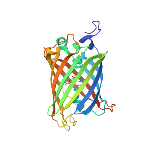
    This study reports the influence of halogens on fluorescence properties of the Aequorea victoria Green Fluorescent Protein variant S65T/T203Y (E(2)GFP). Halide binding forms a specific nonfluorescent complex generating a substantial drop of the fluorescence via static quenching. Spectroscopic analysis under different solution conditions reveals high halogen affinity, which is strongly dependent on the pH. This evidences the presence in E(2)GFP of interacting binding sites for halide ions and for protons. Thermodynamic link and cooperative interaction are assessed demonstrating that binding of one halide ion is associated with the binding of one proton in a cooperative fashion with the formation, in the pH range 4.5-10, of a single fully protonated E(2)GFP.halogen complex. To resolve the structural determinants of E(2)GFP sensitivity to halogens, high-resolution crystallographic structures were obtained for the halide-free and I(-), Br(-), and Cl(-) bound E(2)GFP. Remarkably the first high-resolution (1.4 A) crystallographic structure of a chloride-bound GFP is reported. The chloride ion occupies a specific and unique binding pocket in direct contact (3.4 A) with the chromophore imidazolidinone aromatic ring. Unanticipated flexibility, strongly modulated by halide ion interactions, is observed in the region surrounding the chromophore. Furthermore molecular dynamics simulations identified E222 residue (along with the chromophore Y66 residue) being in the protonated state when E(2)GFP.halogen complex is formed. The impact of these results on high-sensitivity biosensor design will be discussed.


  • Organizational Affiliation
    • Scuola Normale Superiore, National Enterprise for nanoScience and nanoTechnology-Consiglio Nazionale delle Ricerche-Instituto Nazionale di Fisica della Materia, Pisa, Italy. d.arosio@sns.it

Macromolecule Content 

  • Total Structure Weight: 27.54 kDa 
  • Atom Count: 2,148 
  • Modeled Residue Count: 234 
  • Deposited Residue Count: 242 
  • Unique protein chains: 1

Macromolecules

Find similar proteins by:|  3D Structure
Entity ID: 1
MoleculeChains  Sequence LengthOrganismDetailsImage
Green fluorescent protein242Aequorea victoriaMutation(s): 4 
Gene Names: GFP
UniProt
Find proteins for P42212 (Aequorea victoria)
Explore P42212 
Go to UniProtKB:  P42212
Entity Groups
Sequence Clusters30% Identity50% Identity70% Identity90% Identity95% Identity100% Identity
UniProt GroupP42212
Sequence Annotations
Expand
Reference Sequence

Small Molecules

Ligands 1 Unique
IDChains Name / Formula / InChI Key2D Diagram3D Interactions
NA

Query on NA


Download:Ideal Coordinates CCD File
B [auth A]SODIUM ION
Na
FKNQFGJONOIPTF-UHFFFAOYSA-N
Modified Residues  1 Unique
IDChains TypeFormula2D DiagramParent
CRO
Query on CRO
A
L-PEPTIDE LINKINGC15 H17 N3 O5THR, TYR, GLY

Experimental Data & Validation

Experimental Data

  • Method: X-RAY DIFFRACTION
  • Resolution: 1.47 Å
  • R-Value Free:  0.187 (Depositor), 0.193 (DCC) 
  • R-Value Work:  0.159 (Depositor), 0.168 (DCC) 
  • R-Value Observed: 0.160 (Depositor) 
Space Group: P 21 21 21
Unit Cell:
Length ( Å )Angle ( ˚ )
a = 51.133α = 90
b = 62.85β = 90
c = 69.631γ = 90
Software Package:
Software NamePurpose
REFMACrefinement
ADSCdata collection
MOSFLMdata reduction
MOLREPphasing

Structure Validation

View Full Validation Report


Entry History 

Deposition Data

  • Released Date: 2007-05-15 
  • Deposition Author(s): Garau, G.

Revision History  (Full details and data files)

  • Version 1.0: 2007-05-15
    Type: Initial release
  • Version 1.1: 2008-05-01
    Changes: Version format compliance
  • Version 1.2: 2011-07-13
    Changes: Version format compliance
  • Version 1.3: 2018-01-31
    Changes: Experimental preparation
  • Version 1.4: 2021-10-20
    Changes: Database references, Derived calculations
  • Version 1.5: 2024-10-30
    Changes: Data collection, Structure summary
  • Version 2.0: 2026-03-18
    Changes: Polymer sequence