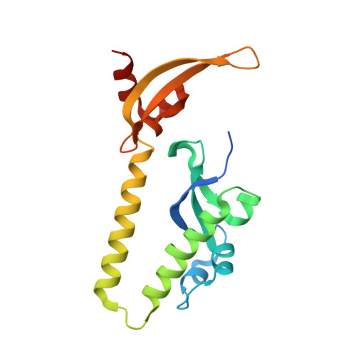
2H1E

Tandem chromodomains of budding yeast CHD1


Experimental Data Snapshot

  • Method: X-RAY DIFFRACTION
  • Resolution: 2.20 Å
  • R-Value Free: 0.246 
  • R-Value Work: 0.189 
  • R-Value Observed: 0.192 

Starting Model: experimental
View more details

wwPDB Validation   3D Report Full Report


This is version 1.4 of the entry. See complete history


Literature

Molecular Implications of Evolutionary Differences in CHD Double Chromodomains.

Flanagan, J.F.Blus, B.J.Kim, D.Clines, K.L.Rastinejad, F.Khorasanizadeh, S.

(2007) J Mol Biol 369: 334-342

  • DOI: https://doi.org/10.1016/j.jmb.2007.03.024
  • Primary Citation of Related Structures:  
    2H1E

  • PubMed Abstract: 

    Double chromodomains occur in CHD proteins, which are ATP-dependent chromatin remodeling factors implicated in RNA polymerase II transcription regulation. Biochemical studies suggest important differences in the histone H3 tail binding of different CHD chromodomains. In human and Drosophila, CHD1 double chromodomains bind lysine 4-methylated histone H3 tail, which is a hallmark of transcriptionally active chromatin in all eukaryotes. Here, we present the crystal structure of the yeast CHD1 double chromodomains, and pinpoint their differences with that of the human CHD1 double chromodomains. The most conserved residues in these double chromodomains are the two chromoboxes that orient adjacently. Only a subset of CHD chromoboxes can form an aromatic cage for methyllysine binding, and methyllysine binding requires correctly oriented inserts. These factors preclude yeast CHD1 double chromodomains from interacting with the histone H3 tail. Despite great sequence similarity between the human CHD1 and CHD2 chromodomains, variation within an insert likely prevents CHD2 double chromodomains from binding lysine 4-methylated histone H3 tail as efficiently as in CHD1. By using the available structural and biochemical data we highlight the evolutionary specialization of CHD double chromodomains, and provide insights about their targeting capacities.


  • Organizational Affiliation

    Department of Biochemistry and Molecular Genetics, University of Virginia Health System, Charlottesville, VA 22908, USA.


Macromolecules
Find similar proteins by:  (by identity cutoff)  |  3D Structure
Entity ID: 1
MoleculeChains Sequence LengthOrganismDetailsImage
Chromo domain protein 1
A, B
177Saccharomyces cerevisiaeMutation(s): 0 
Gene Names: CHD1
EC: 3.6.1 (PDB Primary Data), 3.6.4 (UniProt)
UniProt
Find proteins for P32657 (Saccharomyces cerevisiae (strain ATCC 204508 / S288c))
Explore P32657 
Go to UniProtKB:  P32657
Entity Groups  
Sequence Clusters30% Identity50% Identity70% Identity90% Identity95% Identity100% Identity
UniProt GroupP32657
Sequence Annotations
Expand
  • Reference Sequence
Experimental Data & Validation

Experimental Data

  • Method: X-RAY DIFFRACTION
  • Resolution: 2.20 Å
  • R-Value Free: 0.246 
  • R-Value Work: 0.189 
  • R-Value Observed: 0.192 
  • Space Group: I 41
Unit Cell:
Length ( Å )Angle ( ˚ )
a = 136.185α = 90
b = 136.185β = 90
c = 57.516γ = 90
Software Package:
Software NamePurpose
DENZOdata reduction
SCALEPACKdata scaling
MOLREPphasing
REFMACrefinement
PDB_EXTRACTdata extraction

Structure Validation

View Full Validation Report


Entry History 

Deposition Data

Revision History  (Full details and data files)

  • Version 1.0: 2007-03-27
    Type: Initial release
  • Version 1.1: 2008-05-01
    Changes: Version format compliance
  • Version 1.2: 2011-07-13
    Changes: Advisory, Refinement description, Version format compliance
  • Version 1.3: 2017-10-18
    Changes: Refinement description
  • Version 1.4: 2023-08-30
    Changes: Data collection, Database references, Refinement description