2H03 | pdb_00002h03

Structural studies of protein tyrosine phosphatase beta catalytic domain in complex with inhibitors


Experimental Data Snapshot

  • Method: X-RAY DIFFRACTION
  • Resolution: 1.65 Å
  • R-Value Free: 
    0.217 (Depositor), 0.228 (DCC) 
  • R-Value Work: 
    0.180 (Depositor), 0.189 (DCC) 
  • R-Value Observed: 
    0.180 (Depositor) 

Starting Model: experimental
View more details

wwPDB Validation 3D Report Full Report

Validation slider image for 2H03

Ligand Structure Quality Assessment 


This is version 1.5 of the entry. See complete history

Literature

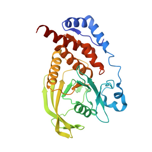
Design and synthesis of potent, non-peptidic inhibitors of HPTPbeta.

Amarasinghe, K.K.Evdokimov, A.G.Xu, K.Clark, C.M.Maier, M.B.Srivastava, A.Colson, A.O.Gerwe, G.S.Stake, G.E.Howard, B.W.Pokross, M.E.Gray, J.L.Peters, K.G.

(2006) Bioorg Med Chem Lett 16: 4252-4256

  • DOI: https://doi.org/10.1016/j.bmcl.2006.05.074
  • Primary Citation Related Structures: 
    2H02, 2H03, 2H04

  • PubMed Abstract: 

    The sulfamic acid phosphotyrosine mimetic was coupled with a previously known malonate template to obtain highly selective and potent inhibitors of HPTPbeta. Potentially hydrolyzable malonate ester functionalities were replaced with 1,2,4-oxadiazoles without a significant effect on HPTPbeta potency.


  • Organizational Affiliation
    • Procter & Gamble Pharmaceuticals, Health Care Research Center, Mason, OH 45040, USA. Amarasinghe.kk@pg.com

Macromolecule Content 

  • Total Structure Weight: 34.39 kDa 
  • Atom Count: 2,625 
  • Modeled Residue Count: 288 
  • Deposited Residue Count: 291 
  • Unique protein chains: 1

Macromolecules

Find similar proteins by:|  3D Structure
Entity ID: 1
MoleculeChains  Sequence LengthOrganismDetailsImage
Protein tyrosine phosphatase, receptor type, B,291Homo sapiensMutation(s): 2 
Gene Names: PTPRB
EC: 3.1.3.48
UniProt & NIH Common Fund Data Resources
Find proteins for P23467 (Homo sapiens)
Explore P23467 
Go to UniProtKB:  P23467
PHAROS:  P23467
GTEx:  ENSG00000127329 
Entity Groups
Sequence Clusters30% Identity50% Identity70% Identity90% Identity95% Identity100% Identity
UniProt GroupP23467
Sequence Annotations
Expand
Reference Sequence

Experimental Data & Validation

Experimental Data

  • Method: X-RAY DIFFRACTION
  • Resolution: 1.65 Å
  • R-Value Free:  0.217 (Depositor), 0.228 (DCC) 
  • R-Value Work:  0.180 (Depositor), 0.189 (DCC) 
  • R-Value Observed: 0.180 (Depositor) 
Space Group: C 1 2 1
Unit Cell:
Length ( Å )Angle ( ˚ )
a = 112.316α = 90
b = 38.934β = 103.91
c = 66.062γ = 90
Software Package:
Software NamePurpose
REFMACrefinement
PDB_EXTRACTdata extraction
MAR345data collection
HKL-2000data scaling
AMoREphasing

Structure Validation

View Full Validation Report


Ligand Structure Quality Assessment 


Entry History 

Deposition Data

Revision History  (Full details and data files)

  • Version 1.0: 2006-06-13
    Type: Initial release
  • Version 1.1: 2008-05-01
    Changes: Version format compliance
  • Version 1.2: 2011-07-13
    Changes: Derived calculations, Version format compliance
  • Version 1.3: 2017-10-18
    Changes: Refinement description
  • Version 1.4: 2021-10-20
    Changes: Database references, Derived calculations
  • Version 1.5: 2023-08-30
    Changes: Data collection, Refinement description