Location of a potential transport binding site in a sigma class glutathione transferase by x-ray crystallography.
Ji, X., von Rosenvinge, E.C., Johnson, W.W., Armstrong, R.N., Gilliland, G.L.(1996) Proc Natl Acad Sci U S A 93: 8208-8213
- PubMed: 8710848 Search on PubMedSearch on PubMed Central
- DOI: https://doi.org/10.1073/pnas.93.16.8208
- Primary Citation Related Structures:
2GSQ - PubMed Abstract:
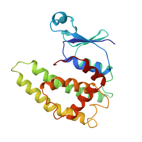
The crystal structure of the sigma class glutathione transferase from squid digestive gland in complex with S-(3-iodobenzyl)glutathione reveals a third binding site for the glutathione conjugate besides the two in the active sites of the dimer. The additional binding site is near the crystallographic two-fold axis between the two alpha 4-turn-alpha 5 motifs. The principal binding interactions with the conjugate include specific electrostatic interactions between the peptide and the two subunits and a hydrophobic cavity found across the two-fold axis that accommodates the 3-iodobenzyl group. Thus, two identical, symmetry-related but mutually exclusive binding modes for the third conjugate are observed. The hydrophobic pocket is about 14 A from the hydroxyl group of Tyr-7 in the active site. This site is a potential transport binding site for hydrophobic molecules or their glutathione conjugates.
- Center for Advanced Research in Biotechnology, University of Maryland Biotechnology Institute, Rockville, USA.
Organizational Affiliation: