2GPZ | pdb_00002gpz

Transthyretin-like protein from Salmonella dublin


Experimental Data Snapshot

  • Method: X-RAY DIFFRACTION
  • Resolution: 2.50 Å
  • R-Value Free: 
    0.261 (Depositor), 0.237 (DCC) 
  • R-Value Work: 
    0.226 (Depositor), 0.237 (DCC) 
  • R-Value Observed: 
    0.228 (Depositor) 

Starting Model: experimental
View more details

wwPDB Validation 3D Report Full Report

Validation slider image for 2GPZ

This is version 1.3 of the entry. See complete history

Literature

The Crystal Structure of the Transthyretin-like Protein from Salmonella dublin, a Prokaryote 5-Hydroxyisourate Hydrolase.

Hennebry, S.C.Law, R.H.Richardson, S.J.Buckle, A.M.Whisstock, J.C.

(2006) J Mol Biology 359: 1389-1399

  • DOI: https://doi.org/10.1016/j.jmb.2006.04.057
  • Primary Citation Related Structures: 
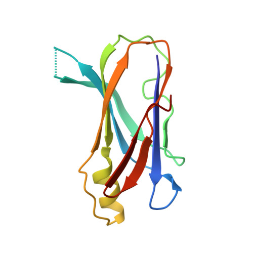
    2GPZ

  • PubMed Abstract: 

    The mechanism of binding of thyroid hormones by the transport protein transthyretin (TTR) in vertebrates is structurally well characterised. However, a homologous family of transthyretin-like proteins (TLPs) present in bacteria as well as eukaryotes do not bind thyroid hormones, instead they are postulated to perform a role in the purine degradation pathway and function as 5-hydroxyisourate hydrolases. Here we describe the 2.5 Angstroms X-ray crystal structure of the TLP from the Gram-negative bacterium Salmonella dublin, and compare and contrast its structure with vertebrate TTRs. The overall architecture of the homotetramer is conserved and, despite low sequence homology with vertebrate TTRs, structural differences within the monomer are restricted to flexible loop regions. However, sequence variation at the dimer-dimer interface has profound consequences for the ligand binding site and provides a structural rationalisation for the absence of thyroid hormone binding affinity in bacterial TLPs: the deep, negatively charged thyroxine-binding pocket that characterises vertebrate TTR contrasts with a shallow and elongated, positively charged cleft in S. dublin TLP. We have demonstrated that Sdu_TLP is a 5-hydroxyisourate hydrolase. Furthermore, using site-directed mutagenesis, we have identified three conserved residues located in this cleft that are critical to the enzyme activity. Together our data reveal that the active site of Sdu_TLP corresponds to the thyroxine binding site in TTRs.


  • Organizational Affiliation
    • The Department of Biochemistry and Molecular Biology, Bio21 Molecular Science and Biotechnology Institute, The University of Melbourne, Parkville, VIC, Australia.

Macromolecule Content 

  • Total Structure Weight: 25.52 kDa 
  • Atom Count: 1,774 
  • Modeled Residue Count: 220 
  • Deposited Residue Count: 222 
  • Unique protein chains: 1

Macromolecules

Find similar proteins by:|  3D Structure
Entity ID: 1
MoleculeChains  Sequence LengthOrganismDetailsImage
transthyretin-like proteinA,
B [auth C]
111Salmonella enterica subsp. enterica serovar DublinMutation(s): 0 
EC: 3.5.2.17
UniProt
Find proteins for Q4VYA5 (Salmonella dublin)
Explore Q4VYA5 
Go to UniProtKB:  Q4VYA5
Entity Groups
Sequence Clusters30% Identity50% Identity70% Identity90% Identity95% Identity100% Identity
UniProt GroupQ4VYA5
Sequence Annotations
Expand
Reference Sequence

Experimental Data & Validation

Experimental Data

  • Method: X-RAY DIFFRACTION
  • Resolution: 2.50 Å
  • R-Value Free:  0.261 (Depositor), 0.237 (DCC) 
  • R-Value Work:  0.226 (Depositor), 0.237 (DCC) 
  • R-Value Observed: 0.228 (Depositor) 
Space Group: P 6
Unit Cell:
Length ( Å )Angle ( ˚ )
a = 94.705α = 90
b = 94.705β = 90
c = 57.381γ = 120
Software Package:
Software NamePurpose
SCALAdata scaling
PHASERphasing
REFMACrefinement
PDB_EXTRACTdata extraction
MOSFLMdata reduction
CCP4data scaling

Structure Validation

View Full Validation Report


Entry History 

Deposition Data

Revision History  (Full details and data files)

  • Version 1.0: 2006-05-16
    Type: Initial release
  • Version 1.1: 2008-05-01
    Changes: Version format compliance
  • Version 1.2: 2011-07-13
    Changes: Version format compliance
  • Version 1.3: 2023-08-30
    Changes: Data collection, Database references, Derived calculations, Refinement description