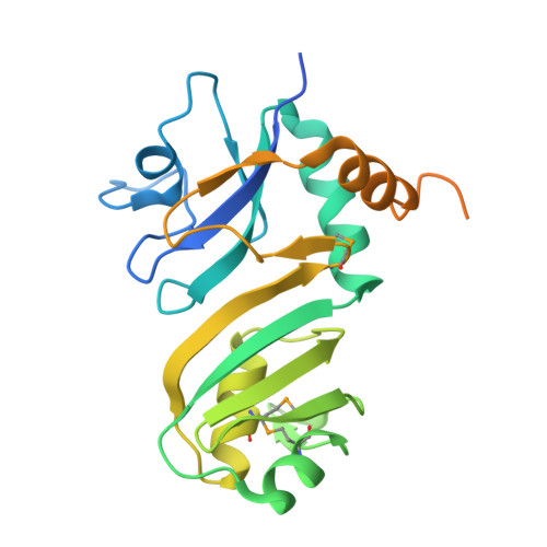
2GML

Crystal Structure of Catalytic Domain of E.coli RluF


Experimental Data Snapshot

  • Method: X-RAY DIFFRACTION
  • Resolution: 2.60 Å
  • R-Value Free: 0.287 
  • R-Value Work: 0.225 

wwPDB Validation   3D Report Full Report


This is version 1.2 of the entry. See complete history


Literature

Domain Organization and Crystal Structure of the Catalytic Domain of E.coli RluF, a Pseudouridine Synthase that Acts on 23S rRNA

Sunita, S.Zhenxing, H.Swaathi, J.Cygler, M.Matte, A.Sivaraman, J.

(2006) J Mol Biol 359: 998-1009

  • DOI: https://doi.org/10.1016/j.jmb.2006.04.019
  • Primary Citation of Related Structures:  
    2GML

  • PubMed Abstract: 

    Pseudouridine synthases catalyze the isomerization of uridine to pseudouridine (Psi) in rRNA and tRNA. The pseudouridine synthase RluF from Escherichia coli (E.C. 4.2.1.70) modifies U2604 in 23S rRNA, and belongs to a large family of pseudouridine synthases present in all kingdoms of life. Here we report the domain architecture and crystal structure of the catalytic domain of E.coli RluF at 2.6A resolution. Limited proteolysis, mass spectrometry and N-terminal sequencing indicate that RluF has a distinct domain architecture, with the catalytic domain flanked at the N and C termini by additional domains connected to it by flexible linkers. The structure of the catalytic domain of RluF is similar to those of RsuA and TruB. RluF is a member of the RsuA sequence family of Psi-synthases, along with RluB and RluE. Structural comparison of RluF with its closest structural homologues, RsuA and TruB, suggests possible functional roles for the N-terminal and C-terminal domains of RluF.


  • Organizational Affiliation

    Department of Biological Sciences, National University of Singapore, 14 Science Drive, Singapore, Singapore 117543.


Macromolecules
Find similar proteins by:  (by identity cutoff)  |  3D Structure
Entity ID: 1
MoleculeChains Sequence LengthOrganismDetailsImage
Ribosomal large subunit pseudouridine synthase F
A, B
237Escherichia coliMutation(s): 4 
EC: 5.4.99 (PDB Primary Data), 5.4.99.21 (UniProt)
UniProt
Find proteins for P32684 (Escherichia coli (strain K12))
Explore P32684 
Go to UniProtKB:  P32684
Entity Groups  
Sequence Clusters30% Identity50% Identity70% Identity90% Identity95% Identity100% Identity
UniProt GroupP32684
Sequence Annotations
Expand
  • Reference Sequence
Small Molecules
Modified Residues  1 Unique
IDChains TypeFormula2D DiagramParent
MSE
Query on MSE
A, B
L-PEPTIDE LINKINGC5 H11 N O2 SeMET
Experimental Data & Validation

Experimental Data

  • Method: X-RAY DIFFRACTION
  • Resolution: 2.60 Å
  • R-Value Free: 0.287 
  • R-Value Work: 0.225 
  • Space Group: P 43 21 2
Unit Cell:
Length ( Å )Angle ( ˚ )
a = 65.714α = 90
b = 65.714β = 90
c = 215.914γ = 90
Software Package:
Software NamePurpose
ADSCdata collection
DENZOdata reduction
SOLVEphasing
CNSrefinement
SCALEPACKdata scaling

Structure Validation

View Full Validation Report


Entry History 

Deposition Data

Revision History  (Full details and data files)

  • Version 1.0: 2006-07-18
    Type: Initial release
  • Version 1.1: 2008-05-01
    Changes: Version format compliance
  • Version 1.2: 2011-07-13
    Changes: Version format compliance