2GM3 | pdb_00002gm3

Crystal Structure of an Universal Stress Protein Family Protein from Arabidopsis Thaliana At3g01520 with AMP Bound


Experimental Data Snapshot

  • Method: X-RAY DIFFRACTION
  • Resolution: 2.46 Å
  • R-Value Free: 
    0.261 (Depositor), 0.260 (DCC) 
  • R-Value Work: 
    0.218 (Depositor), 0.225 (DCC) 
  • R-Value Observed: 
    0.221 (Depositor) 

wwPDB Validation 3D Report Full Report

Validation slider image for 2GM3

Ligand Structure Quality Assessment 


This is version 1.5 of the entry. See complete history

Literature

Crystal structure of the protein At3g01520, a eukaryotic universal stress protein-like protein from arabidopsis thaliana in complex with AMP.

Kim, D.O.J.Bitto, E.Bingman, C.A.Kim, H.J.Han, B.W.Phillips, G.N.

(2015) Proteins 83: 1368-1373

  • DOI: https://doi.org/10.1002/prot.24821
  • Primary Citation Related Structures: 
    2GM3

  • PubMed Abstract: 

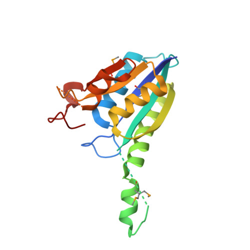
    Members of the universal stress protein (USP) family are conserved in a phylogenetically diverse range of prokaryotes, fungi, protists, and plants and confer abilities to respond to a wide range of environmental stresses. Arabidopsis thaliana contains 44 USP domain-containing proteins, and USP domain is found either in a small protein with unknown physiological function or in an N-terminal portion of a multi-domain protein, usually a protein kinase. Here, we report the first crystal structure of a eukaryotic USP-like protein encoded from the gene At3g01520. The crystal structure of the protein At3g01520 was determined by the single-wavelength anomalous dispersion method and refined to an R factor of 21.8% (Rfree = 26.1%) at 2.5 Å resolution. The crystal structure includes three At3g01520 protein dimers with one AMP molecule bound to each protomer, comprising a Rossmann-like α/β overall fold. The bound AMP and conservation of residues in the ATP-binding loop suggest that the protein At3g01520 also belongs to the ATP-binding USP subfamily members.


  • Organizational Affiliation
    • Research Institute of Pharmaceutical Sciences, College of Pharmacy, Seoul National University, Seoul, 151-742, Korea.

Macromolecule Content 

  • Total Structure Weight: 120.23 kDa 
  • Atom Count: 7,245 
  • Modeled Residue Count: 905 
  • Deposited Residue Count: 1,050 
  • Unique protein chains: 1

Macromolecules

Find similar proteins by:|  3D Structure
Entity ID: 1
MoleculeChains  Sequence LengthOrganismDetailsImage
unknown protein
A, B, C, D, E
A, B, C, D, E, F
175Arabidopsis thalianaMutation(s): 3 
Gene Names: At3g01520
UniProt
Find proteins for Q8LGG8 (Arabidopsis thaliana)
Explore Q8LGG8 
Go to UniProtKB:  Q8LGG8
Entity Groups
Sequence Clusters30% Identity50% Identity70% Identity90% Identity95% Identity100% Identity
UniProt GroupQ8LGG8
Sequence Annotations
Expand
Reference Sequence

Experimental Data & Validation

Experimental Data

  • Method: X-RAY DIFFRACTION
  • Resolution: 2.46 Å
  • R-Value Free:  0.261 (Depositor), 0.260 (DCC) 
  • R-Value Work:  0.218 (Depositor), 0.225 (DCC) 
  • R-Value Observed: 0.221 (Depositor) 
Space Group: P 1
Unit Cell:
Length ( Å )Angle ( ˚ )
a = 63.351α = 75.45
b = 65.662β = 75.04
c = 73.014γ = 66.11
Software Package:
Software NamePurpose
DENZOdata reduction
SCALEPACKdata scaling
RESOLVEphasing
REFMACrefinement
PDB_EXTRACTdata extraction
SHELXEmodel building

Structure Validation

View Full Validation Report


Ligand Structure Quality Assessment 


Entry History 

Deposition Data

Revision History  (Full details and data files)

  • Version 1.0: 2006-04-18
    Type: Initial release
  • Version 1.1: 2008-05-01
    Changes: Version format compliance
  • Version 1.2: 2011-07-13
    Changes: Advisory, Version format compliance
  • Version 1.3: 2016-06-08
    Changes: Database references
  • Version 1.4: 2017-10-18
    Changes: Refinement description
  • Version 1.5: 2024-10-16
    Changes: Data collection, Database references, Derived calculations, Refinement description, Structure summary