2GHM | pdb_00002ghm

Mutated MAP kinase P38 (Mus Musculus) in complex with Inhbitor PG-895449


Experimental Data Snapshot

  • Method: X-RAY DIFFRACTION
  • Resolution: 2.35 Å
  • R-Value Free: 
    0.323 (Depositor) 
  • R-Value Work: 
    0.227 (Depositor), 0.231 (DCC) 
  • R-Value Observed: 
    0.232 (Depositor) 

wwPDB Validation 3D Report Full Report

Validation slider image for 2GHM

Ligand Structure Quality Assessment 


This is version 1.5 of the entry. See complete history. 

Literature

Development of N-2,4-pyrimidine-N-phenyl-N'-phenyl ureas as inhibitors of tumor necrosis factor alpha (TNF-alpha) synthesis. Part 1.

Brugel, T.A., Maier, J.A., Clark, M.P., Sabat, M., Golebiowski, A., Bookland, R.G., Laufersweiler, M.J., Laughlin, S.K., Vanrens, J.C., De, B., Hsieh, L.C., Mekel, M.J., Janusz, M.J.

(2006) Bioorg Med Chem Lett 16: 3510-3513

  • DOI: https://doi.org/10.1016/j.bmcl.2006.03.095
  • Primary Citation Related Structures: 
    2GHL, 2GHM

  • PubMed Abstract: 

    A new class of tumor necrosis factor alpha (TNF-alpha) synthesis inhibitors based on an N-2,4-pyrimidine-N-phenyl-N'-phenyl urea scaffold is described. Many of these compounds showed low-nanomolar activity against lipopolysaccharide stimulated TNF-alpha production. X-ray co-crystallization studies with mutated p38alpha showed that these trisubstituted ureas interact with the ATP-binding pocket in a pseudo-bicyclic conformation brought about by the presence of an intramolecular hydrogen bonding interaction.


  • Organizational Affiliation: 
    • Procter and Gamble Pharmaceuticals, Health Care Research Center, 8700 Mason-Montgomery Rd., Mason, OH 45040, USA. brugel.ta@pg.com

Macromolecule Content 

  • Total Structure Weight: 40.32 kDa 
  • Atom Count: 2,867 
  • Modeled Residue Count: 336 
  • Deposited Residue Count: 348 
  • Unique protein chains: 1

Macromolecules

Find similar proteins by:|  3D Structure
Entity ID: 1
MoleculeChains  Sequence LengthOrganismDetailsImage
Mitogen-activated protein kinase 14348Mus musculusMutation(s): 2 
Gene Names: Mapk14, Crk1, Csbp1, Csbp2
EC: 2.7.1.37 (PDB Primary Data), 2.7.11.24 (UniProt)
UniProt
Find proteins for P47811 (Mus musculus)
Explore P47811 
Go to UniProtKB:  P47811
Entity Groups
Sequence Clusters30% Identity50% Identity70% Identity90% Identity95% Identity100% Identity
UniProt GroupP47811
Sequence Annotations
Expand
Reference Sequence

Small Molecules

Ligands 1 Unique
IDChains Name / Formula / InChI Key2D Diagram3D Interactions
LIC

Query on LIC


Download:Ideal Coordinates CCD File
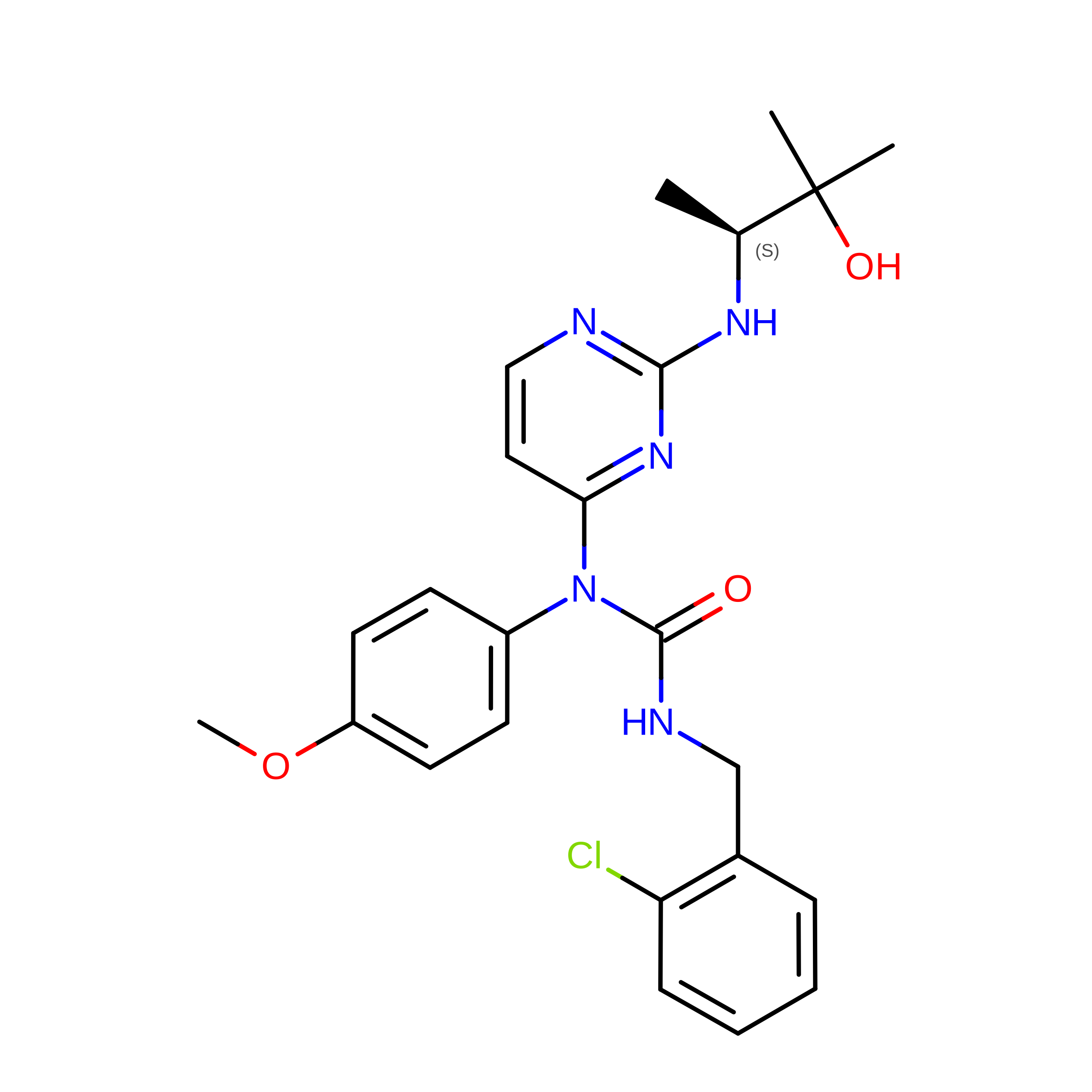
B [auth A]3-(2-CHLOROBENZYL)-1-(2-{[(1S)-2-HYDROXY-1,2-DIMETHYLPROPYL]AMINO}PYRIMIDIN-4-YL)-1-(4-METHOXYPHENYL)UREA
C24 H28 Cl N5 O3
ZHBOBAJCKHPQBT-INIZCTEOSA-N

Experimental Data & Validation

Experimental Data

  • Method: X-RAY DIFFRACTION
  • Resolution: 2.35 Å
  • R-Value Free:  0.323 (Depositor) 
  • R-Value Work:  0.227 (Depositor), 0.231 (DCC) 
  • R-Value Observed: 0.232 (Depositor) 
Space Group: P 21 21 21
Unit Cell:
Length ( Å )Angle ( ˚ )
a = 65.169α = 90
b = 74.855β = 90
c = 77.879γ = 90
Software Package:
Software NamePurpose
DENZOdata reduction
SCALEPACKdata scaling
AMoREphasing
REFMACrefinement
PDB_EXTRACTdata extraction
MAR345data collection

Structure Validation

View Full Validation Report


Ligand Structure Quality Assessment 


Entry History 

Deposition Data

Revision History  (Full details and data files)

  • Version 1.0: 2006-05-02
    Type: Initial release
  • Version 1.1: 2008-05-01
    Changes: Version format compliance
  • Version 1.2: 2011-07-13
    Changes: Version format compliance
  • Version 1.3: 2017-10-18
    Changes: Refinement description
  • Version 1.4: 2021-10-20
    Changes: Database references, Derived calculations
  • Version 1.5: 2024-02-14
    Changes: Data collection