


Structure of hypothetical protein ph0006 from Pyrococcus horikoshii
Cuff, M.E., Skarina, T., Gorodichtchenskaia, E., Edwards, A., Savchenko, A., Joachimiak, A.To be published.
Experimental Data Snapshot
wwPDB Validation 3D Report Full Report

Entity ID: 1 | |||||
|---|---|---|---|---|---|
| Molecule | Chains | Sequence Length | Organism | Details | Image |
| UPF0204 protein PH0006 | 298 | Pyrococcus horikoshii OT3 | Mutation(s): 9 Gene Names: PH0006 EC: 3.1.1.96 | 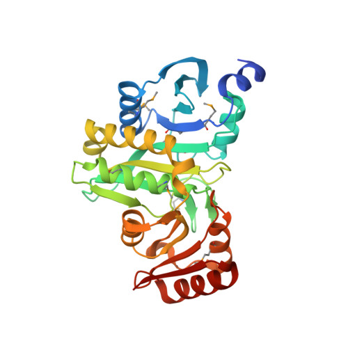 | |
UniProt | |||||
Entity Groups | |||||
| Sequence Clusters | 30% Identity50% Identity70% Identity90% Identity95% Identity100% Identity | ||||
| UniProt Group | O57774 | ||||
Sequence AnnotationsExpand | |||||
Reference Sequence | |||||
| Ligands 2 Unique | |||||
|---|---|---|---|---|---|
| ID | Chains | Name / Formula / InChI Key | 2D Diagram | 3D Interactions | |
| SO4 Download:Ideal Coordinates CCD File | D [auth A] E [auth A] F [auth A] G [auth A] H [auth A] | SULFATE ION O4 S QAOWNCQODCNURD-UHFFFAOYSA-L |  | ||
| MG Download:Ideal Coordinates CCD File | J [auth A], R [auth B], W [auth C] | MAGNESIUM ION Mg JLVVSXFLKOJNIY-UHFFFAOYSA-N |  | ||
| Modified Residues 1 Unique | |||||
|---|---|---|---|---|---|
| ID | Chains | Type | Formula | 2D Diagram | Parent |
| MSE Query on MSE | A, B, C | L-PEPTIDE LINKING | C5 H11 N O2 Se |  | MET |
| Length ( Å ) | Angle ( ˚ ) |
|---|---|
| a = 65.021 | α = 90 |
| b = 67.029 | β = 90.09 |
| c = 111.572 | γ = 90 |
| Software Name | Purpose |
|---|---|
| REFMAC | refinement |
| SBC-Collect | data collection |
| HKL-2000 | data scaling |
| HKL-3000 | phasing |
| SHELX | phasing |
| MLPHARE | phasing |
| DM | phasing |
| SOLVE | phasing |
| RESOLVE | phasing |
| Coot | model building |
| O | model building |
| ARP/wARP | model building |


