2GFJ | pdb_00002gfj

Crystal structure of the zinc-beta-lactamase L1 from stenotrophomonas maltophilia (inhibitor 1)


Experimental Data Snapshot

  • Method: X-RAY DIFFRACTION
  • Resolution: 1.80 Å
  • R-Value Free: 
    0.239 (Depositor), 0.239 (DCC) 
  • R-Value Work: 
    0.196 (Depositor), 0.196 (DCC) 
  • R-Value Observed: 
    0.198 (Depositor) 

Starting Model: experimental
View more details

wwPDB Validation 3D Report Full Report

Validation slider image for 2GFJ

Ligand Structure Quality Assessment 


This is version 1.5 of the entry. See complete history

Literature

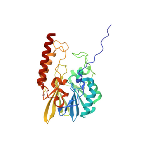
Structural insights into the design of inhibitors for the L1 metallo-beta-lactamase from Stenotrophomonas maltophilia.

Nauton, L.Kahn, R.Garau, G.Hernandez, J.F.Dideberg, O.

(2008) J Mol Biology 375: 257-269

  • DOI: https://doi.org/10.1016/j.jmb.2007.10.036
  • Primary Citation Related Structures: 
    2FM6, 2FU7, 2FU8, 2FU9, 2GFJ, 2GFK, 2H6A, 2HB9

  • PubMed Abstract: 

    One mechanism by which bacteria can escape the action of beta-lactam antibiotics is the production of metallo-beta-lactamases. Inhibition of these enzymes should restore the action of these widely used antibiotics. The tetrameric enzyme L1 from Stenotrophomonas maltophilia was used as a model system to determine a series of high-resolution crystal structures of apo, mono and bi-metal substituted proteins as well as protein-inhibitor complexes. Unexpectedly, although the apo structure revealed only few significant structural differences from the holo structure, some inhibitors were shown to induce amino acid side-chain rotations in the tightly packed active site. Moreover, one inhibitor employs a new binding mode in order to interact with the di-zinc center. This structural information could prove essential in the process of elucidation of the mode of interaction between a putative lead compound and metallo-beta-lactamases, one of the main steps in structure-based drug design.


  • Organizational Affiliation
    • Institut de Biologie Structurale Jean-Pierre Ebel (UMR 5075;CNRS;CEA;UJF), 41, rue Jules Horowitz, F-38027 Grenoble Cedex 1, France.

Macromolecule Content 

  • Total Structure Weight: 59.13 kDa 
  • Atom Count: 4,579 
  • Modeled Residue Count: 532 
  • Deposited Residue Count: 538 
  • Unique protein chains: 1

Macromolecules

Find similar proteins by:|  3D Structure
Entity ID: 1
MoleculeChains  Sequence LengthOrganismDetailsImage
Metallo-beta-lactamase L1
A, B
269Stenotrophomonas maltophiliaMutation(s): 0 
Gene Names: L1
EC: 3.5.2.6
UniProt
Find proteins for P52700 (Stenotrophomonas maltophilia)
Explore P52700 
Go to UniProtKB:  P52700
Entity Groups
Sequence Clusters30% Identity50% Identity70% Identity90% Identity95% Identity100% Identity
UniProt GroupP52700
Sequence Annotations
Expand
Reference Sequence

Small Molecules

Ligands 3 Unique
IDChains Name / Formula / InChI Key2D Diagram3D Interactions
VI

Query on VI


Download:Ideal Coordinates CCD File
I [auth A],
P [auth B]
1,3-DIPHENYL-1H-PYRAZOLE-4,5-DICARBOXYLIC ACID
C17 H12 N2 O4
WERNQSNSNVDBBJ-UHFFFAOYSA-N
SO4

Query on SO4


Download:Ideal Coordinates CCD File
E [auth A]
F [auth A]
G [auth A]
H [auth A]
L [auth B]
E [auth A],
F [auth A],
G [auth A],
H [auth A],
L [auth B],
M [auth B],
N [auth B],
O [auth B]
SULFATE ION
O4 S
QAOWNCQODCNURD-UHFFFAOYSA-L
ZN

Query on ZN


Download:Ideal Coordinates CCD File
C [auth A],
D [auth A],
J [auth B],
K [auth B]
ZINC ION
Zn
PTFCDOFLOPIGGS-UHFFFAOYSA-N

Experimental Data & Validation

Experimental Data

  • Method: X-RAY DIFFRACTION
  • Resolution: 1.80 Å
  • R-Value Free:  0.239 (Depositor), 0.239 (DCC) 
  • R-Value Work:  0.196 (Depositor), 0.196 (DCC) 
  • R-Value Observed: 0.198 (Depositor) 
Space Group: P 62 2 2
Unit Cell:
Length ( Å )Angle ( ˚ )
a = 105.88α = 90
b = 105.88β = 90
c = 197.86γ = 120
Software Package:
Software NamePurpose
XDSdata scaling
SCALAdata scaling
CCP4model building
REFMACrefinement
XDSdata reduction
CCP4data scaling
CCP4phasing

Structure Validation

View Full Validation Report


Ligand Structure Quality Assessment 


Entry History 

Deposition Data

Revision History  (Full details and data files)

  • Version 1.0: 2007-03-27
    Type: Initial release
  • Version 1.1: 2008-04-01
    Changes: Version format compliance
  • Version 1.2: 2011-07-13
    Changes: Version format compliance
  • Version 1.3: 2017-10-18
    Changes: Refinement description
  • Version 1.4: 2023-10-25
    Changes: Data collection, Database references, Derived calculations, Refinement description
  • Version 1.5: 2024-10-23
    Changes: Structure summary