2GCL | pdb_00002gcl

Structure of the Pob3 Middle domain


Experimental Data Snapshot

  • Method: X-RAY DIFFRACTION
  • Resolution: 2.21 Å
  • R-Value Free: 
    0.264 (Depositor), 0.264 (DCC) 
  • R-Value Work: 
    0.207 (Depositor), 0.210 (DCC) 
  • R-Value Observed: 
    0.210 (Depositor) 

wwPDB Validation 3D Report Full Report

Validation slider image for 2GCL

This is version 1.4 of the entry. See complete history

Literature

The Structure of the yFACT Pob3-M Domain, Its Interaction with the DNA Replication Factor RPA, and a Potential Role in Nucleosome Deposition.

VanDemark, A.P.Blanksma, M.Ferris, E.Heroux, A.Hill, C.P.Formosa, T.

(2006) Mol Cell 22: 363-374

  • DOI: https://doi.org/10.1016/j.molcel.2006.03.025
  • Primary Citation Related Structures: 
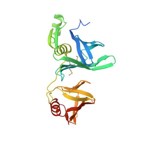
    2GCJ, 2GCL

  • PubMed Abstract: 

    We report the crystal structure of the middle domain of the Pob3 subunit (Pob3-M) of S. cerevisiae FACT (yFACT, facilitates chromatin transcription), which unexpectedly adopts an unusual double pleckstrin homology (PH) architecture. A mutation within a conserved surface cluster in this domain causes a defect in DNA replication that is suppressed by mutation of replication protein A (RPA). The nucleosome reorganizer yFACT therefore interacts in a physiologically important way with the central single-strand DNA (ssDNA) binding factor RPA to promote a step in DNA replication. Purified yFACT and RPA display a weak direct physical interaction, although the genetic suppression is not explained by simple changes in affinity between the purified proteins. Further genetic analysis suggests that coordinated function by yFACT and RPA is important during nucleosome deposition. These results support the model that the FACT family has an essential role in constructing nucleosomes during DNA replication, and suggest that RPA contributes to this process.


  • Organizational Affiliation
    • Department of Biochemistry, University of Utah School of Medicine, Salt Lake City, 84132, USA.

Macromolecule Content 

  • Total Structure Weight: 61.4 kDa 
  • Atom Count: 4,050 
  • Modeled Residue Count: 461 
  • Deposited Residue Count: 522 
  • Unique protein chains: 1

Macromolecules

Find similar proteins by:|  3D Structure
Entity ID: 1
MoleculeChains  Sequence LengthOrganismDetailsImage
Hypothetical 63.0 kDa protein in DAK1-ORC1 intergenic region
A, B
261Saccharomyces cerevisiaeMutation(s): 5 
UniProt
Find proteins for Q04636 (Saccharomyces cerevisiae (strain ATCC 204508 / S288c))
Explore Q04636 
Go to UniProtKB:  Q04636
Entity Groups
Sequence Clusters30% Identity50% Identity70% Identity90% Identity95% Identity100% Identity
UniProt GroupQ04636
Sequence Annotations
Expand
Reference Sequence

Experimental Data & Validation

Experimental Data

  • Method: X-RAY DIFFRACTION
  • Resolution: 2.21 Å
  • R-Value Free:  0.264 (Depositor), 0.264 (DCC) 
  • R-Value Work:  0.207 (Depositor), 0.210 (DCC) 
  • R-Value Observed: 0.210 (Depositor) 
Space Group: P 21 21 21
Unit Cell:
Length ( Å )Angle ( ˚ )
a = 57.11α = 90
b = 57.77β = 90
c = 156.58γ = 90
Software Package:
Software NamePurpose
REFMACrefinement
ADSCdata collection
DENZOdata reduction
SCALEPACKdata scaling
SOLVEphasing

Structure Validation

View Full Validation Report


Entry History 

Deposition Data

Revision History  (Full details and data files)

  • Version 1.0: 2006-05-23
    Type: Initial release
  • Version 1.1: 2008-05-01
    Changes: Version format compliance
  • Version 1.2: 2011-07-13
    Changes: Version format compliance
  • Version 1.3: 2021-10-20
    Changes: Database references, Derived calculations
  • Version 1.4: 2024-10-30
    Changes: Data collection, Structure summary