2GCE | pdb_00002gce

The 1,1-proton transfer reaction mechanism by alpha-methylacyl-CoA racemase is catalyzed by an aspartate/histidine pair and involves a smooth, methionine-rich surface for binding the fatty acyl moiety


Experimental Data Snapshot

  • Method: X-RAY DIFFRACTION
  • Resolution: 1.85 Å
  • R-Value Free: 
    0.246 (Depositor), 0.247 (DCC) 
  • R-Value Work: 
    0.202 (Depositor), 0.227 (DCC) 
  • R-Value Observed: 
    0.202 (Depositor) 

Starting Model: experimental
View more details

wwPDB Validation 3D Report Full Report

Validation slider image for 2GCE

Ligand Structure Quality Assessment 


This is version 1.4 of the entry. See complete history

Literature

The Catalysis of the 1,1-Proton Transfer by alpha-Methyl-acyl-CoA Racemase Is Coupled to a Movement of the Fatty Acyl Moiety Over a Hydrophobic, Methionine-rich Surface

Bhaumik, P.Schmitz, W.Hassinen, A.Hiltunen, J.K.Conzelmann, E.Wierenga, R.K.

(2007) J Mol Biology 367: 1145-1161

  • DOI: https://doi.org/10.1016/j.jmb.2007.01.062
  • Primary Citation Related Structures: 
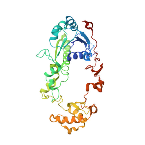
    2GCE, 2GCI, 2GD0, 2GD2, 2GD6

  • PubMed Abstract: 

    Alpha-methylacyl-CoA racemases are essential enzymes for branched-chain fatty acid metabolism. Their reaction mechanism and the structural basis of their wide substrate specificity are poorly understood. High-resolution crystal structures of Mycobacterium tuberculosis alpha-methylacyl-CoA racemase (MCR) complexed with substrate molecules show the active site geometry required for catalysis of the interconversion of (2S) and (2R)-methylacyl-CoA. The thioester oxygen atom and the 2-methyl group are in a cis-conformation with respect to each other. The thioester oxygen atom fits into an oxyanion hole and the 2-methyl group points into a hydrophobic pocket. The active site geometry agrees with a 1,1-proton transfer mechanism in which the acid/base-pair residues are His126 and Asp156. The structures of the complexes indicate that the acyl chains of the S-substrate and the R-substrate bind in an S-pocket and an R-pocket, respectively. A unique feature of MCR is a large number of methionine residues in the acyl binding region, located between the S-pocket and the R-pocket. It appears that the (S) to (R) interconversion of the 2-methylacyl chiral center is coupled to a movement of the acyl group over this hydrophobic, methionine-rich surface, when moving from its S-pocket to its R-pocket, whereas the 2-methyl moiety and the CoA group remain fixed in their respective pockets.


  • Organizational Affiliation
    • Biocenter Oulu and Department of Biochemistry, University of Oulu, Linnanmaa, P.O. Box 3000, FIN-90014 University of Oulu, Finland.

Macromolecule Content 

  • Total Structure Weight: 162.55 kDa 
  • Atom Count: 12,350 
  • Modeled Residue Count: 1,416 
  • Deposited Residue Count: 1,440 
  • Unique protein chains: 1

Macromolecules

Find similar proteins by:|  3D Structure
Entity ID: 1
MoleculeChains  Sequence LengthOrganismDetailsImage
probable alpha-methylacyl-CoA racemase MCR
A, B, C, D
360Mycobacterium tuberculosisMutation(s): 0 
Gene Names: CAB09031
EC: 5.1.99.4
UniProt
Find proteins for O06543 (Mycobacterium tuberculosis (strain ATCC 25618 / H37Rv))
Explore O06543 
Go to UniProtKB:  O06543
Entity Groups
Sequence Clusters30% Identity50% Identity70% Identity90% Identity95% Identity100% Identity
UniProt GroupO06543
Sequence Annotations
Expand
Reference Sequence

Small Molecules

Ligands 2 Unique
IDChains Name / Formula / InChI Key2D Diagram3D Interactions
RFC

Query on RFC


Download:Ideal Coordinates CCD File
F [auth A],
H [auth B],
J [auth C],
L [auth D]
(R)-IBUPROFENOYL-COENZYME A
C34 H53 N7 O17 P3 S
NXIKDQUQQHUCJH-NGSKWOHTSA-O
SFC

Query on SFC


Download:Ideal Coordinates CCD File
E [auth A],
G [auth B],
I [auth C],
K [auth D]
(S)-IBUPROFENOYL-COENZYME A
C34 H53 N7 O17 P3 S
NXIKDQUQQHUCJH-MIXAKNBRSA-O

Experimental Data & Validation

Experimental Data

  • Method: X-RAY DIFFRACTION
  • Resolution: 1.85 Å
  • R-Value Free:  0.246 (Depositor), 0.247 (DCC) 
  • R-Value Work:  0.202 (Depositor), 0.227 (DCC) 
  • R-Value Observed: 0.202 (Depositor) 
Space Group: C 1 2 1
Unit Cell:
Length ( Å )Angle ( ˚ )
a = 179.95α = 90
b = 79.58β = 90.07
c = 117.43γ = 90
Software Package:
Software NamePurpose
REFMACrefinement
MAR345data collection
XDSdata scaling
MOLREPphasing

Structure Validation

View Full Validation Report


Ligand Structure Quality Assessment 


Entry History 

Deposition Data

Revision History  (Full details and data files)

  • Version 1.0: 2007-02-20
    Type: Initial release
  • Version 1.1: 2008-05-01
    Changes: Version format compliance
  • Version 1.2: 2011-07-13
    Changes: Version format compliance
  • Version 1.3: 2017-10-18
    Changes: Refinement description
  • Version 1.4: 2023-10-25
    Changes: Data collection, Database references, Derived calculations, Refinement description, Structure summary