2G9Q | pdb_00002g9q

The crystal structure of the glycogen phosphorylase b- 1AB complex


Experimental Data Snapshot

  • Method: X-RAY DIFFRACTION
  • Resolution: 2.50 Å
  • R-Value Free: 
    0.243 (Depositor) 
  • R-Value Work: 
    0.190 (Depositor) 
  • R-Value Observed: 
    0.192 (Depositor) 

Starting Model: experimental
View more details

wwPDB Validation 3D Report Full Report

Validation slider image for 2G9Q

This is version 1.4 of the entry. See complete history

Literature

Iminosugars as potential inhibitors of glycogenolysis: structural insights into the molecular basis of glycogen phosphorylase inhibition.

Oikonomakos, N.G.Tiraidis, C.Leonidas, D.D.Zographos, S.E.Kristiansen, M.Jessen, C.U.Norskov-Lauritsen, L.Agius, L.

(2006) J Med Chem 49: 5687-5701

  • DOI: https://doi.org/10.1021/jm060496g
  • Primary Citation Related Structures: 
    2G9Q, 2G9R, 2G9U, 2G9V

  • PubMed Abstract: 

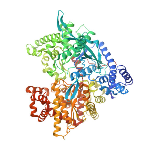
    Iminosugars DAB (5), isofagomine (9), and several N-substituted derivatives have been identified as potent inhibitors of liver glycogen phosphorylase a (IC(50) = 0.4-1.2 microM) and of basal and glucagon-stimulated glycogenolysis (IC(50) = 1-3 microM). The X-ray structures of 5, 9, and its N-3-phenylpropyl analogue 8 in complex with rabbit muscle glycogen phosphorylase (GPb) shows that iminosugars bind tightly at the catalytic site in the presence of the substrate phosphate and induce conformational changes that characterize the R-state conformation of the enzyme. Charged nitrogen N1 is within hydrogen-bonding distance with the carbonyl oxygen of His377 (5) and in ionic contact with the substrate phosphate oxygen (8 and 9). Our findings suggest that the inhibitors function as oxocarbenium ion transition-state analogues. The conformational change to the R state provides an explanation for previous findings that 5, unlike inhibitors that favor the T state, promotes phosphorylation of GPb in hepatocytes with sequential inactivation of glycogen synthase.


  • Organizational Affiliation
    • Institute of Organic and Pharmaceutical Chemistry, The National Hellenic Research Foundation, Athens 11635, Greece. ngo@eie.gr

Macromolecule Content 

  • Total Structure Weight: 97.94 kDa 
  • Atom Count: 6,664 
  • Modeled Residue Count: 804 
  • Deposited Residue Count: 842 
  • Unique protein chains: 1

Macromolecules

Find similar proteins by:|  3D Structure
Entity ID: 1
MoleculeChains  Sequence LengthOrganismDetailsImage
Glycogen phosphorylase, muscle form842Oryctolagus cuniculusMutation(s): 1 
EC: 2.4.1.1
UniProt
Find proteins for P00489 (Oryctolagus cuniculus)
Explore P00489 
Go to UniProtKB:  P00489
Entity Groups
Sequence Clusters30% Identity50% Identity70% Identity90% Identity95% Identity100% Identity
UniProt GroupP00489
Sequence Annotations
Expand
Reference Sequence

Small Molecules

Modified Residues  1 Unique
IDChains TypeFormula2D DiagramParent
LLP
Query on LLP
A
L-PEPTIDE LINKINGC14 H22 N3 O7 PLYS

Experimental Data & Validation

Experimental Data

  • Method: X-RAY DIFFRACTION
  • Resolution: 2.50 Å
  • R-Value Free:  0.243 (Depositor) 
  • R-Value Work:  0.190 (Depositor) 
  • R-Value Observed: 0.192 (Depositor) 
Space Group: P 43 21 2
Unit Cell:
Length ( Å )Angle ( ˚ )
a = 128.712α = 90
b = 128.712β = 90
c = 117.442γ = 90
Software Package:
Software NamePurpose
REFMACrefinement
DENZOdata reduction
SCALEPACKdata scaling

Structure Validation

View Full Validation Report


Entry History 

Deposition Data

Revision History  (Full details and data files)

  • Version 1.0: 2007-01-16
    Type: Initial release
  • Version 1.1: 2008-05-01
    Changes: Version format compliance
  • Version 1.2: 2011-07-13
    Changes: Version format compliance
  • Version 1.3: 2023-08-30
    Changes: Data collection, Database references, Derived calculations, Refinement description
  • Version 1.4: 2023-11-15
    Changes: Data collection