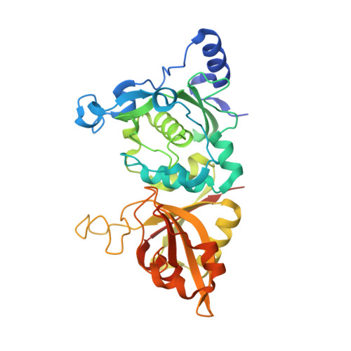
2G8J

Calpain 1 proteolytic core in complex with SNJ-1945, a alpha-ketoamide-type inhibitor.


Experimental Data Snapshot

  • Method: X-RAY DIFFRACTION
  • Resolution: 1.61 Å
  • R-Value Free: 0.229 
  • R-Value Work: 0.197 
  • R-Value Observed: 0.198 

Starting Model: experimental
View more details

wwPDB Validation   3D Report Full Report


Ligand Structure Quality Assessment 


This is version 1.5 of the entry. See complete history


Literature

Calpain Inhibition by alpha-Ketoamide and Cyclic Hemiacetal Inhibitors Revealed by X-ray Crystallography

Cuerrier, D.Moldoveanu, T.Inoue, J.Davies, P.L.Campbell, R.L.

(2006) Biochemistry 45: 7446-7452

  • DOI: https://doi.org/10.1021/bi060425j
  • Primary Citation of Related Structures:  
    2G8E, 2G8J

  • PubMed Abstract: 

    Calpains are intracellular calcium-activated cysteine proteases whose unregulated proteolysis following the loss of calcium homeostasis can lead to acute degeneration during ischemic episodes and trauma, as well as Alzheimer's disease and cataract formation. The determination of the crystal structure of the proteolytic core of mu-calpain (muI-II) in a calcium-bound active conformation has made structure-guided design of active site inhibitors feasible. We present here high-resolution crystal structures of rat muI-II complexed with two reversible calpain-specific inhibitors employing cyclic hemiacetal (SNJ-1715) and alpha-ketoamide (SNJ-1945) chemistries that reveal new details about the interactions of inhibitors with this enzyme. The SNJ-1715 complex confirms that the free aldehyde is the reactive species of the cornea-permeable cyclic hemiacetal. The alpha-ketoamide warhead of SNJ-1945 binds with the hydroxyl group of the tetrahedral adduct pointing toward the catalytic histidine rather than the oxyanion hole. The muI-II-SNJ-1945 complex shows residue Glu261 displaced from the S1' site by the inhibitor, resulting in an extended "open" conformation of the domain II gating loop and an unobstructed S1' site. This conformation offers an additional template for structure-based drug design extending to the primed subsites. An important role for the highly conserved Glu261 is proposed.


  • Organizational Affiliation

    Department of Biochemistry, Queen's University, Kingston, Ontario, Canada K7L 3N6.


Macromolecules
Find similar proteins by:  (by identity cutoff)  |  3D Structure
Entity ID: 1
MoleculeChains Sequence LengthOrganismDetailsImage
Calpain-1 catalytic subunit339Rattus norvegicusMutation(s): 0 
Gene Names: Capn1Cls1
EC: 3.4.22.52
UniProt
Find proteins for P97571 (Rattus norvegicus)
Explore P97571 
Go to UniProtKB:  P97571
Entity Groups  
Sequence Clusters30% Identity50% Identity70% Identity90% Identity95% Identity100% Identity
UniProt GroupP97571
Sequence Annotations
Expand
  • Reference Sequence
Small Molecules
Binding Affinity Annotations 
IDSourceBinding Affinity
D7G BindingDB:  2G8J IC50: 170 (nM) from 1 assay(s)
Experimental Data & Validation

Experimental Data

  • Method: X-RAY DIFFRACTION
  • Resolution: 1.61 Å
  • R-Value Free: 0.229 
  • R-Value Work: 0.197 
  • R-Value Observed: 0.198 
  • Space Group: P 21 21 21
Unit Cell:
Length ( Å )Angle ( ˚ )
a = 40.572α = 90
b = 70.4β = 90
c = 110.895γ = 90
Software Package:
Software NamePurpose
SCALAdata scaling
MOLREPphasing
REFMACrefinement
PDB_EXTRACTdata extraction
MAR345data collection
CCP4data scaling

Structure Validation

View Full Validation Report


Ligand Structure Quality Assessment 


Entry History 

Deposition Data

Revision History  (Full details and data files)

  • Version 1.0: 2006-06-06
    Type: Initial release
  • Version 1.1: 2008-05-01
    Changes: Version format compliance
  • Version 1.2: 2011-07-13
    Changes: Version format compliance
  • Version 1.3: 2017-10-18
    Changes: Refinement description
  • Version 1.4: 2023-08-30
    Changes: Data collection, Database references, Derived calculations, Refinement description
  • Version 1.5: 2024-10-30
    Changes: Structure summary