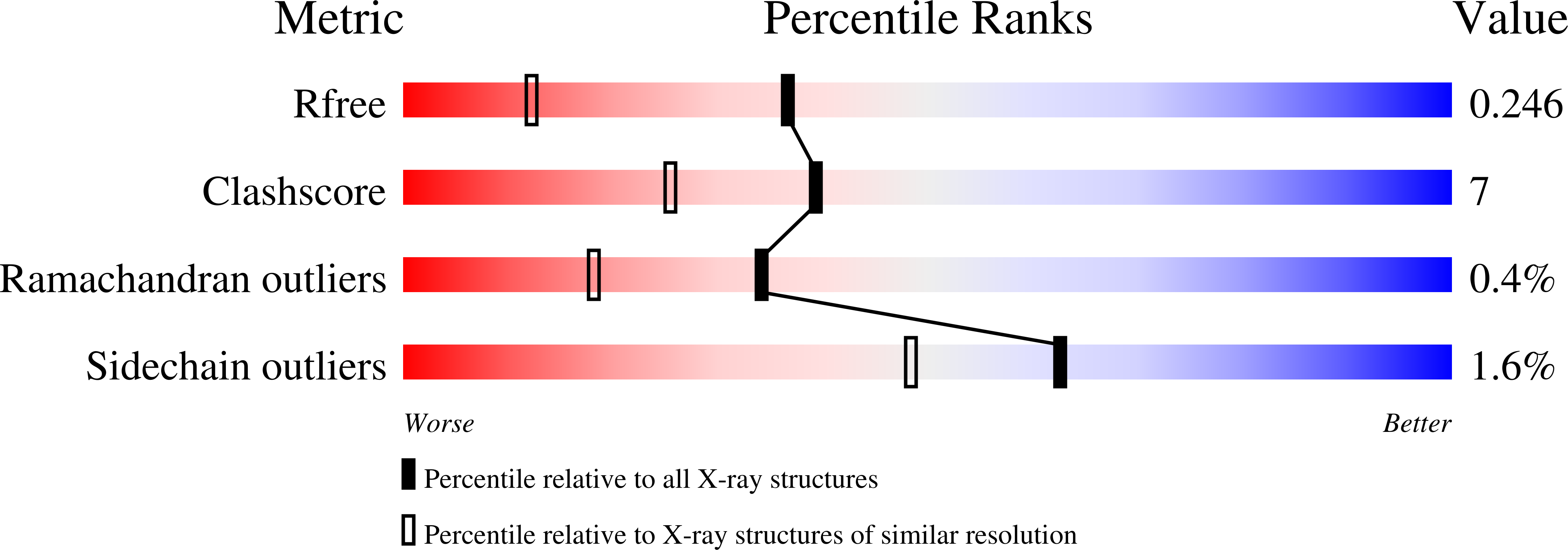
2FZV

Crystal Structure of an apo form of a Flavin-binding Protein from Shigella flexneri


Experimental Data Snapshot

  • Method: X-RAY DIFFRACTION
  • Resolution: 1.70 Å
  • R-Value Free: 0.246 
  • R-Value Work: 0.199 
  • R-Value Observed: 0.201 

wwPDB Validation   3D Report Full Report


This is version 1.4 of the entry. See complete history


Literature

Crystal structure of an apo form of Shigella flexneri ArsH protein with an NADPH-dependent FMN reductase activity

Vorontsov, I.I.Minasov, G.Brunzelle, J.S.Shuvalova, L.Kiryukhina, O.Collart, F.R.Anderson, W.F.

(2007) Protein Sci 16: 2483-2490

  • DOI: https://doi.org/10.1110/ps.073029607
  • Primary Citation of Related Structures:  
    2FZV

  • PubMed Abstract: 

    The arsH gene or its homologs are a frequent part of the arsenic resistance system in bacteria and eukaryotes. Although a specific biological function of the gene product is unknown, the ArsH protein was annotated as a member of the NADPH-dependent FMN reductase family based on a conserved (T/S)XRXXSX(T/S) fingerprint motif common for FMN binding proteins. Presented here are the first crystal structure of an ArsH protein from Shigella flexneri refined at 1.7 A resolution and results of enzymatic activity assays that revealed a strong NADPH-dependent FMN reductase and low azoreductase activities. The ArsH apo protein has an alpha/beta/alpha-fold typical for FMN binding proteins. The asymmetric unit consists of four monomers, which form a tetramer. Buried surface analysis suggests that this tetramer is likely to be the relevant biological assembly. Dynamic light scattering experiments are consistent with this hypothesis and show that ArsH in solution at room temperature does exist predominantly in the tetrameric form.


  • Organizational Affiliation

    Department of Molecular Pharmacology and Biological Chemistry, Northwestern University, Feinberg School of Medicine, Chicago, IL 60611, USA.


Macromolecules
Find similar proteins by:  (by identity cutoff)  |  3D Structure
Entity ID: 1
MoleculeChains Sequence LengthOrganismDetailsImage
putative arsenical resistance protein
A, B, C, D
279Shigella flexneri 2a str. 2457TMutation(s): 0 
Gene Names: AAP17869
EC: 1
UniProt
Find proteins for Q7UC03 (Shigella flexneri)
Explore Q7UC03 
Go to UniProtKB:  Q7UC03
Entity Groups  
Sequence Clusters30% Identity50% Identity70% Identity90% Identity95% Identity100% Identity
UniProt GroupQ7UC03
Sequence Annotations
Expand
  • Reference Sequence
Experimental Data & Validation

Experimental Data

  • Method: X-RAY DIFFRACTION
  • Resolution: 1.70 Å
  • R-Value Free: 0.246 
  • R-Value Work: 0.199 
  • R-Value Observed: 0.201 
  • Space Group: P 43 21 2
Unit Cell:
Length ( Å )Angle ( ˚ )
a = 117.821α = 90
b = 117.821β = 90
c = 154.08γ = 90
Software Package:
Software NamePurpose
REFMACrefinement
MAR345data collection
XDSdata scaling
SCALEPACKdata scaling
SHARPphasing

Structure Validation

View Full Validation Report


Entry History 

Revision History  (Full details and data files)

  • Version 1.0: 2006-02-21
    Type: Initial release
  • Version 1.1: 2008-05-01
    Changes: Version format compliance
  • Version 1.2: 2011-07-13
    Changes: Advisory, Source and taxonomy, Version format compliance
  • Version 1.3: 2017-10-18
    Changes: Refinement description
  • Version 1.4: 2024-02-14
    Changes: Data collection, Database references, Derived calculations