2FZS | pdb_00002fzs

Crystal structure of E. coli ClpP with a Peptide Chloromethyl Ketone Covalently Bound at the Active Site


Experimental Data Snapshot

  • Method: X-RAY DIFFRACTION
  • Resolution: 1.90 Å
  • R-Value Free: 
    0.233 (Depositor), 0.322 (DCC) 
  • R-Value Work: 
    0.173 (Depositor) 
  • R-Value Observed: 
    0.176 (Depositor) 

Starting Model: experimental
View more details

wwPDB Validation 3D Report Full Report

Validation slider image for 2FZS

Ligand Structure Quality Assessment 


This is version 1.4 of the entry. See complete history

Literature

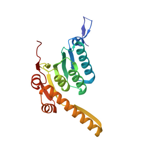
Crystal structure at 1.9A of E. coli ClpP with a peptide covalently bound at the active site.

Szyk, A.Maurizi, M.R.

(2006) J Struct Biol 156: 165-174

  • DOI: https://doi.org/10.1016/j.jsb.2006.03.013
  • Primary Citation Related Structures: 
    2FZS

  • PubMed Abstract: 

    ClpP, the proteolytic component of the ATP-dependent ClpAP and ClpXP chaperone/protease complexes, has 14 identical subunits organized in two stacked heptameric rings. The active sites are in an interior aqueous chamber accessible through axial channels. We have determined a 1.9 A crystal structure of Escherichia coli ClpP with benzyloxycarbonyl-leucyltyrosine chloromethyl ketone (Z-LY-CMK) bound at each active site. The complex mimics a tetrahedral intermediate during peptide cleavage, with the inhibitor covalently linked to the active site residues, Ser97 and His122. Binding is further stabilized by six hydrogen bonds between backbone atoms of the peptide and ClpP as well as by hydrophobic binding of the phenolic ring of tyrosine in the S1 pocket. The peptide portion of Z-LY-CMK displaces three water molecules in the native enzyme resulting in little change in the conformation of the peptide binding groove. The heptameric rings of ClpP-CMK are slightly more compact than in native ClpP, but overall structural changes were minimal (rmsd approximately 0.5 A). The side chain of Ser97 is rotated approximately 90 degrees in forming the covalent adduct with Z-LY-CMK, indicating that rearrangement of the active site residues to a active configuration occurs upon substrate binding. The N-terminal peptide of ClpP-CMK is stabilized in a beta-hairpin conformation with the proximal N-terminal residues lining the axial channel and the loop extending beyond the apical surface of the heptameric ring. The lack of major substrate-induced conformational changes suggests that changes in ClpP structure needed to facilitate substrate entry or product release must be limited to rigid body motions affecting subunit packing or contacts between ClpP rings.


  • Organizational Affiliation
    • Laboratory of Cell Biology, National Cancer Institute, National Institutes of Health, Bethesda, MD 20892, USA.

Macromolecule Content 

  • Total Structure Weight: 311.48 kDa 
  • Atom Count: 24,125 
  • Modeled Residue Count: 2,591 
  • Deposited Residue Count: 2,702 
  • Unique protein chains: 1

Macromolecules

Find similar proteins by:|  3D Structure
Entity ID: 1
MoleculeChains  Sequence LengthOrganismDetailsImage
ATP-dependent Clp protease proteolytic subunit
A, B, C, D, E
A, B, C, D, E, F, G, H, I, J, K, L, M, N
193Escherichia coliMutation(s): 0 
Gene Names: clpPlopP
EC: 3.4.21.92
UniProt
Find proteins for P0A6G7 (Escherichia coli (strain K12))
Explore P0A6G7 
Go to UniProtKB:  P0A6G7
Entity Groups
Sequence Clusters30% Identity50% Identity70% Identity90% Identity95% Identity100% Identity
UniProt GroupP0A6G7
Sequence Annotations
Expand
Reference Sequence

Small Molecules

Ligands 3 Unique
IDChains Name / Formula / InChI Key2D Diagram3D Interactions
CMQ

Query on CMQ


Download:Ideal Coordinates CCD File
BA [auth E]
DA [auth F]
GA [auth G]
JA [auth H]
MA [auth I]
BA [auth E],
DA [auth F],
GA [auth G],
JA [auth H],
MA [auth I],
O [auth A],
OA [auth J],
QA [auth K],
R [auth B],
UA [auth L],
V [auth C],
XA [auth M],
Z [auth D],
ZA [auth N]
N~2~-[(BENZYLOXY)CARBONYL]-N-[(1S,2S)-2-HYDROXY-1-(4-HYDROXYBENZYL)PROPYL]-L-LEUCINAMIDE
C24 H32 N2 O5
ZUWYQZGBCBSHFK-HSQYWUDLSA-N
PGE

Query on PGE


Download:Ideal Coordinates CCD File
AA [auth D]
AB [auth N]
CA [auth E]
EA [auth F]
HA [auth G]
AA [auth D],
AB [auth N],
CA [auth E],
EA [auth F],
HA [auth G],
KA [auth H],
NA [auth I],
P [auth A],
PA [auth J],
RA [auth K],
S [auth B],
SA [auth K],
VA [auth L],
W [auth C],
YA [auth M]
TRIETHYLENE GLYCOL
C6 H14 O4
ZIBGPFATKBEMQZ-UHFFFAOYSA-N
GOL

Query on GOL


Download:Ideal Coordinates CCD File
BB [auth N]
FA [auth F]
IA [auth G]
LA [auth H]
Q [auth A]
BB [auth N],
FA [auth F],
IA [auth G],
LA [auth H],
Q [auth A],
T [auth B],
TA [auth K],
U [auth B],
WA [auth L],
X [auth C],
Y [auth C]
GLYCEROL
C3 H8 O3
PEDCQBHIVMGVHV-UHFFFAOYSA-N

Experimental Data & Validation

Experimental Data

  • Method: X-RAY DIFFRACTION
  • Resolution: 1.90 Å
  • R-Value Free:  0.233 (Depositor), 0.322 (DCC) 
  • R-Value Work:  0.173 (Depositor) 
  • R-Value Observed: 0.176 (Depositor) 
Space Group: C 1 2 1
Unit Cell:
Length ( Å )Angle ( ˚ )
a = 190.7α = 90
b = 101β = 99
c = 155.4γ = 90
Software Package:
Software NamePurpose
REFMACrefinement
HKL-2000data reduction
SCALEPACKdata scaling
AMoREphasing

Structure Validation

View Full Validation Report


Ligand Structure Quality Assessment 


Entry History 

Deposition Data

Revision History  (Full details and data files)

  • Version 1.0: 2006-05-23
    Type: Initial release
  • Version 1.1: 2008-05-01
    Changes: Version format compliance
  • Version 1.2: 2011-07-13
    Changes: Advisory, Version format compliance
  • Version 1.3: 2023-08-30
    Changes: Data collection, Database references, Derived calculations, Refinement description
  • Version 1.4: 2024-10-30
    Changes: Structure summary