2FUZ | pdb_00002fuz

UGL hexagonal crystal structure without glycine and DTT molecules


Experimental Data Snapshot

  • Method: X-RAY DIFFRACTION
  • Resolution: 1.80 Å
  • R-Value Free: 
    0.188 (Depositor), 0.194 (DCC) 
  • R-Value Work: 
    0.167 (Depositor), 0.174 (DCC) 
  • R-Value Observed: 
    0.167 (Depositor) 

Starting Model: experimental
View more details

wwPDB Validation 3D Report Full Report

Validation slider image for 2FUZ

This is version 1.3 of the entry. See complete history

Literature

Substrate recognition by unsaturated glucuronyl hydrolase from Bacillus sp. GL1

Itoh, T.Hashimoto, W.Mikami, B.Murata, K.

(2006) Biochem Biophys Res Commun 344: 253-262

  • DOI: https://doi.org/10.1016/j.bbrc.2006.03.141
  • Primary Citation Related Structures: 
    2FUZ, 2FV0, 2FV1

  • PubMed Abstract: 

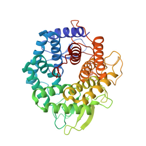
    Bacterial unsaturated glucuronyl hydrolases (UGLs) together with polysaccharide lyases are responsible for the complete depolymerization of mammalian extracellular matrix glycosaminoglycans. UGL acts on various oligosaccharides containing unsaturated glucuronic acid (DeltaGlcA) at the nonreducing terminus and releases DeltaGlcA through hydrolysis. In this study, we demonstrate the substrate recognition mechanism of the UGL of Bacillus sp. GL1 by determining the X-ray crystallographic structure of its substrate-enzyme complexes. The tetrasaccharide-enzyme complex demonstrated that at least four subsites are present in the active pocket. Although several amino acid residues are crucial for substrate binding, the enzyme strongly recognizes DeltaGlcA at subsite -1 through the formation of hydrogen bonds and stacking interactions, and prefers N-acetyl-d-galactosamine and glucose rather than N-acetyl-d-glucosamine as a residue accommodated in subsite +1, due to the steric hindrance.


  • Organizational Affiliation
    • Division of Agronomy and Horticultural Science, Graduate School of Agriculture, Kyoto University, Gokasho, Uji, Kyoto 611-0011, Japan.

Macromolecule Content 

  • Total Structure Weight: 43.98 kDa 
  • Atom Count: 3,604 
  • Modeled Residue Count: 377 
  • Deposited Residue Count: 377 
  • Unique protein chains: 1

Macromolecules

Find similar proteins by:|  3D Structure
Entity ID: 1
MoleculeChains  Sequence LengthOrganismDetailsImage
Unsaturated glucuronyl hydrolase377Bacillus sp. GL1Mutation(s): 0 
Gene Names: ugl
EC: 3.2.1 (PDB Primary Data), 3.2.1.179 (UniProt)
UniProt
Find proteins for Q9RC92 (Bacillus sp. (strain GL1))
Explore Q9RC92 
Go to UniProtKB:  Q9RC92
Entity Groups
Sequence Clusters30% Identity50% Identity70% Identity90% Identity95% Identity100% Identity
UniProt GroupQ9RC92
Sequence Annotations
Expand
Reference Sequence

Small Molecules

Ligands 1 Unique
IDChains Name / Formula / InChI Key2D Diagram3D Interactions
MPD

Query on MPD


Download:Ideal Coordinates CCD File
B [auth A]
C [auth A]
D [auth A]
E [auth A]
F [auth A]
B [auth A],
C [auth A],
D [auth A],
E [auth A],
F [auth A],
G [auth A],
H [auth A],
I [auth A],
J [auth A]
(4S)-2-METHYL-2,4-PENTANEDIOL
C6 H14 O2
SVTBMSDMJJWYQN-YFKPBYRVSA-N

Experimental Data & Validation

Experimental Data

  • Method: X-RAY DIFFRACTION
  • Resolution: 1.80 Å
  • R-Value Free:  0.188 (Depositor), 0.194 (DCC) 
  • R-Value Work:  0.167 (Depositor), 0.174 (DCC) 
  • R-Value Observed: 0.167 (Depositor) 
Space Group: P 65 2 2
Unit Cell:
Length ( Å )Angle ( ˚ )
a = 102.818α = 90
b = 102.818β = 90
c = 223.156γ = 120
Software Package:
Software NamePurpose
CNSrefinement
HKL-2000data reduction
SCALEPACKdata scaling
CNSphasing

Structure Validation

View Full Validation Report


Entry History 

Deposition Data

Revision History  (Full details and data files)

  • Version 1.0: 2006-05-30
    Type: Initial release
  • Version 1.1: 2008-05-01
    Changes: Version format compliance
  • Version 1.2: 2011-07-13
    Changes: Source and taxonomy, Version format compliance
  • Version 1.3: 2023-10-25
    Changes: Data collection, Database references, Derived calculations, Refinement description