2FR4 | pdb_00002fr4

Structure of Fab DNA-1 complexed with a stem-loop DNA ligand


Experimental Data Snapshot

  • Method: X-RAY DIFFRACTION
  • Resolution: 1.95 Å
  • R-Value Free: 
    0.256 (Depositor), 0.261 (DCC) 
  • R-Value Work: 
    0.207 (Depositor), 0.215 (DCC) 
  • R-Value Observed: 
    0.210 (Depositor) 

Starting Model: experimental
View more details

wwPDB Validation 3D Report Full Report

Validation slider image for 2FR4

This is version 1.5 of the entry. See complete history

Literature

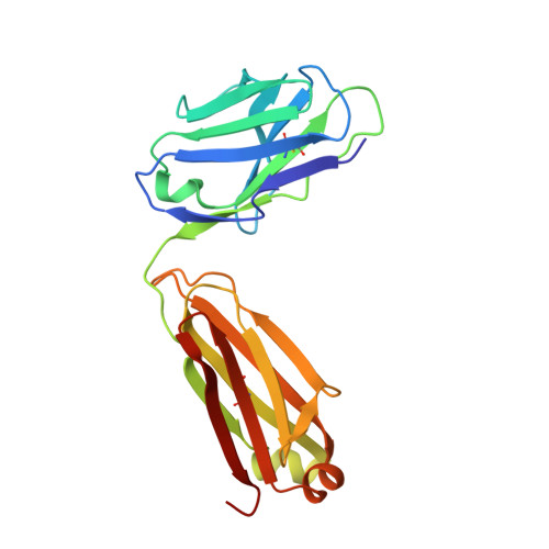
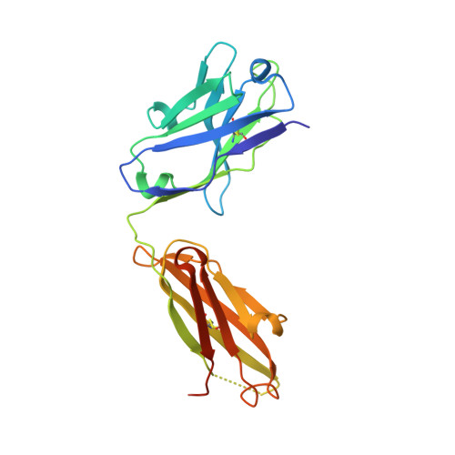
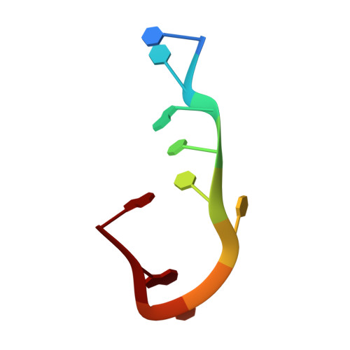
Impact of DNA hairpin folding energetics on antibody-ssDNA association.

Ou, Z.Bottoms, C.A.Henzl, M.T.Tanner, J.J.

(2007) J Mol Biology 374: 1029-1040

  • DOI: https://doi.org/10.1016/j.jmb.2007.09.084
  • Primary Citation Related Structures: 
    2FR4

  • PubMed Abstract: 

    Deposition of anti-DNA antibodies in the kidney contributes to the pathogenesis of the autoimmune disease, systemic lupus erythematosus. Antibodies that bind to hairpin-forming DNA ligands may be particularly prone to deposition. Here we report the first structure of a Fab complexed with hairpin-forming DNA. The ligand used for co-crystallization is 5'-d [CTG(CCTT)CAG]-3', which has a predicted hairpin structure consisting of a four-nucleotide loop (CCTT) and a stem of three base-pairs. The 1.95 A resolution crystal structure of Fab DNA-1 complexed with this ligand shows that the conformation of the bound ligand differs radically from the predicted hairpin conformation. The three base-pairs in the stem are absent in the bound form. The protein binds to the last six nucleotides at the 3' end of the ligand. These nucleotides form a loop (TTCA) closed by a G:C base-pair in the bound state. Stacking of aromatic side-chains against DNA bases is the dominant interaction in the complex. Interactions with the DNA backbone are conspicuously absent. Thermodynamics of binding are examined using isothermal titration calorimetry. The apparent dissociation constant is 4 microM, and binding is enthalpically favorable and entropically unfavorable. Increasing the number of base-pairs in the DNA stem from three to six decreases binding affinity. These data suggest a conformational selection binding mechanism in which the Fab binds preferentially to the unstructured state of the ligand. In this interpretation, the ligand binding and ligand folding equilibria are coupled, with lower hairpin stability leading to greater effective binding affinity. Thus, pre-organization of the DNA loop into the preferred binding conformation does not play a major role in complexation. Rather, it is argued that the stem of the hairpin serves to reduce the degrees of freedom in the free DNA ligand, thereby limiting the entropic cost attendant to complexation with the Fab.


  • Organizational Affiliation
    • Department of Biochemistry, University of Missouri-Columbia, Columbia, MO 65211, USA.

Macromolecule Content 

  • Total Structure Weight: 103.38 kDa 
  • Atom Count: 7,283 
  • Modeled Residue Count: 873 
  • Deposited Residue Count: 908 
  • Unique protein chains: 2
  • Unique nucleic acid chains: 1

Macromolecules


Find similar proteins by:|  3D Structure
Entity ID: 2
MoleculeChains  Sequence LengthOrganismDetailsImage
antibody light chain FABC [auth L],
E [auth A]
214Mus musculusMutation(s): 0 
UniProt
Find proteins for P01837 (Mus musculus)
Explore P01837 
Go to UniProtKB:  P01837
Entity Groups
Sequence Clusters30% Identity50% Identity70% Identity90% Identity95% Identity100% Identity
UniProt GroupP01837
Sequence Annotations
Expand
Reference Sequence
Find similar proteins by:|  3D Structure
Entity ID: 3
MoleculeChains  Sequence LengthOrganismDetailsImage
antibody heavy chain FABD [auth H],
F [auth B]
230Mus musculusMutation(s): 0 
Find similar nucleic acids by:  Sequence
Entity ID: 1
MoleculeChains LengthOrganismImage
5'-D(*CP*TP*GP*CP*CP*TP*TP*CP*AP*G)-3'A [auth M],
B [auth N]
10N/A
Sequence Annotations
Expand
Reference Sequence

Small Molecules

Ligands 1 Unique
IDChains Name / Formula / InChI Key2D Diagram3D Interactions
PG4

Query on PG4


Download:Ideal Coordinates CCD File
G [auth H]TETRAETHYLENE GLYCOL
C8 H18 O5
UWHCKJMYHZGTIT-UHFFFAOYSA-N

Experimental Data & Validation

Experimental Data

  • Method: X-RAY DIFFRACTION
  • Resolution: 1.95 Å
  • R-Value Free:  0.256 (Depositor), 0.261 (DCC) 
  • R-Value Work:  0.207 (Depositor), 0.215 (DCC) 
  • R-Value Observed: 0.210 (Depositor) 
Space Group: P 21 21 21
Unit Cell:
Length ( Å )Angle ( ˚ )
a = 84.757α = 90
b = 90.45β = 90
c = 128.116γ = 90
Software Package:
Software NamePurpose
DENZOdata reduction
SCALEPACKdata scaling
REFMACrefinement
PDB_EXTRACTdata extraction
MOLREPphasing

Structure Validation

View Full Validation Report


Entry History 

Deposition Data

Revision History  (Full details and data files)

  • Version 1.0: 2007-01-09
    Type: Initial release
  • Version 1.1: 2008-05-01
    Changes: Version format compliance
  • Version 1.2: 2011-07-13
    Changes: Advisory, Version format compliance
  • Version 1.3: 2017-10-18
    Changes: Refinement description
  • Version 1.4: 2023-08-30
    Changes: Data collection, Database references, Derived calculations, Refinement description
  • Version 1.5: 2024-11-13
    Changes: Structure summary