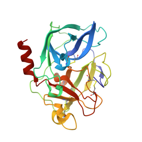
Multiple solvent crystal structures: Probing binding sites, plasticity and hydration
Mattos, C., Bellamacina, C.R., Peisach, E., Pereira, A., Vitkup, D., Petsko, G.A., Ringe, D.(2006) J Mol Biology 357: 1471-1482
- PubMed: 16488429
- DOI: https://doi.org/10.1016/j.jmb.2006.01.039
- Primary Citation of Related Structures:
2FO9, 2FOA, 2FOB, 2FOC, 2FOD, 2FOE, 2FOF, 2FOG, 2FOH - PubMed Abstract:
Multiple solvent crystal structures (MSCS) of porcine pancreatic elastase were used to map the binding surface the enzyme. Crystal structures of elastase in neat acetonitrile, 95% acetone, 55% dimethylformamide, 80% 5-hexene-1,2-diol, 80% isopropanol, 80% ethanol and 40% trifluoroethanol showed that the organic solvent molecules clustered in the active site, were found mostly unclustered in crystal contacts and in general did not bind elsewhere on the surface of elastase. Mixtures of 40% benzene or 40% cyclohexane in 50% isopropanol and 10% water showed no bound benzene or cyclohexane molecules, but did reveal bound isopropanol. The clusters of organic solvent probe molecules coincide with pockets occupied by known inhibitors. MSCS also reveal the areas of plasticity within the elastase binding site and allow for the visualization of a nearly complete first hydration shell. The pattern of organic solvent clusters determined by MSCS for elastase is consistent with patterns for hot spots in protein-ligand interactions determined from database analysis in general. The MSCS method allows probing of hot spots, plasticity and hydration simultaneously, providing a powerful complementary strategy to guide computational methods currently in development for binding site determination, ligand docking and design.
- Department of Molecular and Structural Biochemistry, North Carolina State University, Campus Box 7622, 128 Polk Hall, Raleigh, NC 27695, USA. carla_mattos@ncsu.edu
Organizational Affiliation:


