2FMM

Crystal Structure of EMSY-HP1 complex


Experimental Data Snapshot

  • Method: X-RAY DIFFRACTION
  • Resolution: 1.80 Å
  • R-Value Free: 0.243 
  • R-Value Work: 0.219 
  • R-Value Observed: 0.219 

wwPDB Validation   3D Report Full Report


This is version 1.3 of the entry. See complete history


Literature

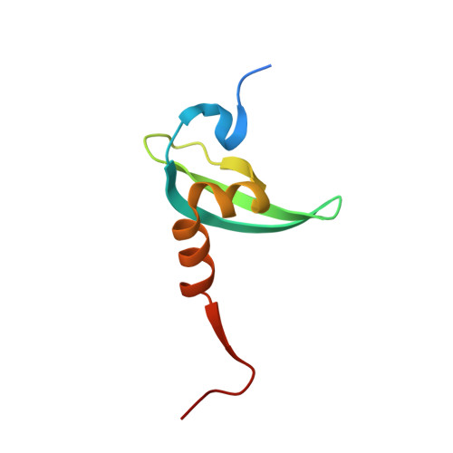
Crystal structure of the HP1-EMSY complex reveals an unusual mode of HP1 binding.

Huang, Y.Myers, M.P.Xu, R.M.

(2006) Structure 14: 703-712

  • DOI: https://doi.org/10.1016/j.str.2006.01.007
  • Primary Citation of Related Structures:  
    2FMM

  • PubMed Abstract: 

    Heterochromatin protein-1 (HP1) plays an essential role in both the assembly of higher-order chromatin structure and epigenetic inheritance. The C-terminal chromo shadow domain (CSD) of HP1 is responsible for homodimerization and interaction with a number of chromatin-associated nonhistone proteins, including EMSY, which is a BRCA2-interacting protein that has been implicated in the development of breast and ovarian cancer. We have determined the crystal structure of the HP1beta CSD in complex with the N-terminal domain of EMSY at 1.8 A resolution. Surprisingly, the structure reveals that EMSY is bound by two HP1 CSD homodimers, and the binding sequences differ from the consensus HP1 binding motif PXVXL. This structural information expands our understanding of HP1 binding specificity and provides insights into interactions between HP1 homodimers that are likely to be important for heterochromatin formation.


  • Organizational Affiliation

    Cold Spring Harbor Laboratory, Cold Spring Harbor, New York 11724, USA.


Macromolecules
Find similar proteins by:  (by identity cutoff)  |  3D Structure
Entity ID: 1
MoleculeChains Sequence LengthOrganismDetailsImage
Protein EMSYA [auth E]133Homo sapiensMutation(s): 0 
Gene Names: EMSYC11orf30
UniProt & NIH Common Fund Data Resources
Find proteins for Q7Z589 (Homo sapiens)
Explore Q7Z589 
Go to UniProtKB:  Q7Z589
PHAROS:  Q7Z589
GTEx:  ENSG00000158636 
Entity Groups  
Sequence Clusters30% Identity50% Identity70% Identity90% Identity95% Identity100% Identity
UniProt GroupQ7Z589
Sequence Annotations
Expand
  • Reference Sequence
Find similar proteins by:  (by identity cutoff)  |  3D Structure
Entity ID: 2
MoleculeChains Sequence LengthOrganismDetailsImage
Chromobox protein homolog 1B [auth A],
C [auth B],
D [auth C],
E [auth D]
74Homo sapiensMutation(s): 0 
Gene Names: CBX1CBX
UniProt & NIH Common Fund Data Resources
Find proteins for P83916 (Homo sapiens)
Explore P83916 
Go to UniProtKB:  P83916
PHAROS:  P83916
GTEx:  ENSG00000108468 
Entity Groups  
Sequence Clusters30% Identity50% Identity70% Identity90% Identity95% Identity100% Identity
UniProt GroupP83916
Sequence Annotations
Expand
  • Reference Sequence
Experimental Data & Validation

Experimental Data

  • Method: X-RAY DIFFRACTION
  • Resolution: 1.80 Å
  • R-Value Free: 0.243 
  • R-Value Work: 0.219 
  • R-Value Observed: 0.219 
  • Space Group: P 21 21 2
Unit Cell:
Length ( Å )Angle ( ˚ )
a = 143.4α = 90
b = 63.87β = 90
c = 66.64γ = 90
Software Package:
Software NamePurpose
HKL-2000data collection
SCALEPACKdata scaling
SOLVEphasing
CNSrefinement
HKL-2000data reduction

Structure Validation

View Full Validation Report


Entry History 

Deposition Data

  • Released Date: 2006-05-23 
  • Deposition Author(s): Huang, Y.

Revision History  (Full details and data files)

  • Version 1.0: 2006-05-23
    Type: Initial release
  • Version 1.1: 2008-05-01
    Changes: Version format compliance
  • Version 1.2: 2011-07-13
    Changes: Derived calculations, Version format compliance
  • Version 1.3: 2024-02-14
    Changes: Data collection, Database references, Derived calculations