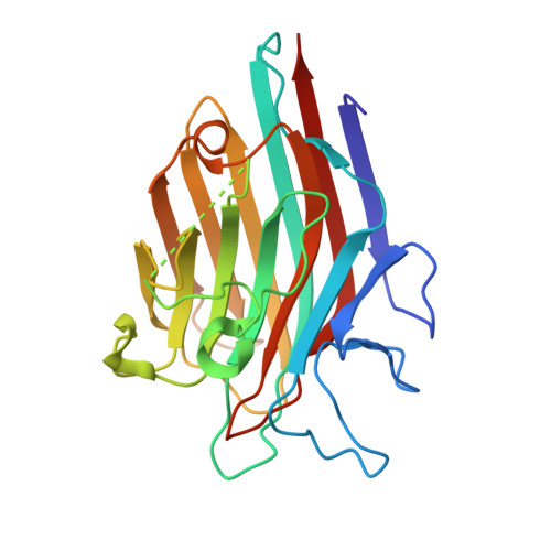
2FMD

Structural basis of carbohydrate recognition by Bowringia milbraedii seed agglutinin


Experimental Data Snapshot

  • Method: X-RAY DIFFRACTION
  • Resolution: 1.90 Å
  • R-Value Free: 0.228 
  • R-Value Work: 0.183 
  • R-Value Observed: 0.186 

Starting Model: experimental
View more details

wwPDB Validation   3D Report Full Report


This is version 2.2 of the entry. See complete history


Literature

Structural basis of carbohydrate recognition by a Man(alpha1-2)Man-specific lectin from Bowringia milbraedii.

Buts, L.Garcia-Pino, A.Wyns, L.Loris, R.

(2006) Glycobiology 16: 635-640

  • DOI: https://doi.org/10.1093/glycob/cwj109
  • Primary Citation of Related Structures:  
    2FMD

  • PubMed Abstract: 

    The crystal structure of the seed lectin from the tropical legume Bowringia milbraedii was determined in complex with the disaccharide ligand Man(alpha1-2)Man. In solution, the protein exhibits a dynamic dimer-tetramer equilibrium, consistent with the concanavalin A-type tetramer observed in the crystal. Contacts between the tetramers are mediated almost exclusively through the carbohydrate ligand, resulting in a crystal lattice virtually identical to that of the concanavalin-A:Man(alpha1-2)Man complex, even though both proteins have less than 50% sequence identity. The disaccharide binds exclusively in a "downstream" binding mode, with the non-reducing mannose occupying the monosaccharide-binding site. The reducing mannose is bound in a predominantly polar subsite involving Tyr131, Gln218, and Tyr219.


  • Organizational Affiliation

    Laboratorium voor Ultrastructuur, Vrije Universiteit Brussel and Department of Molecular and Cellular Interactions, Vlaams Interuniversitair Instituut voor Biotechnologie, Pleinlaan 2, B-1050 Brussels, Belgium. lievbuts@vub.ac.be


Macromolecules
Find similar proteins by:  (by identity cutoff)  |  3D Structure
Entity ID: 1
MoleculeChains Sequence LengthOrganismDetailsImage
Lectin240Leucomphalos mildbraediiMutation(s): 0 
UniProt
Find proteins for P42088 (Leucomphalos mildbraedii)
Explore P42088 
Go to UniProtKB:  P42088
Entity Groups  
Sequence Clusters30% Identity50% Identity70% Identity90% Identity95% Identity100% Identity
UniProt GroupP42088
Sequence Annotations
Expand
  • Reference Sequence
Oligosaccharides

Help

Entity ID: 2
MoleculeChains Length2D Diagram Glycosylation3D Interactions
alpha-D-mannopyranose-(1-2)-alpha-D-mannopyranose
B
2N/A
Glycosylation Resources
GlyTouCan:  G53402KW
GlyCosmos:  G53402KW
GlyGen:  G53402KW
Biologically Interesting Molecules (External Reference) 1 Unique
Experimental Data & Validation

Experimental Data

  • Method: X-RAY DIFFRACTION
  • Resolution: 1.90 Å
  • R-Value Free: 0.228 
  • R-Value Work: 0.183 
  • R-Value Observed: 0.186 
  • Space Group: I 2 2 2
Unit Cell:
Length ( Å )Angle ( ˚ )
a = 66.603α = 90
b = 86.351β = 90
c = 91.76γ = 90
Software Package:
Software NamePurpose
DENZOdata reduction
SCALEPACKdata scaling
PHASERphasing
CNSrefinement

Structure Validation

View Full Validation Report


Entry History 

Deposition Data

Revision History  (Full details and data files)

  • Version 1.0: 2006-08-22
    Type: Initial release
  • Version 1.1: 2008-05-01
    Changes: Version format compliance
  • Version 1.2: 2011-07-13
    Changes: Version format compliance
  • Version 2.0: 2020-07-29
    Type: Remediation
    Reason: Carbohydrate remediation
    Changes: Atomic model, Data collection, Derived calculations, Structure summary
  • Version 2.1: 2023-08-30
    Changes: Data collection, Database references, Refinement description, Structure summary
  • Version 2.2: 2024-11-13
    Changes: Structure summary