2FHE | pdb_00002fhe

FASCIOLA HEPATICA GLUTATHIONE S-TRANSFERASE ISOFORM 1 IN COMPLEX WITH GLUTATHIONE


Experimental Data Snapshot

  • Method: X-RAY DIFFRACTION
  • Resolution: 2.30 Å
  • R-Value Free: 
    0.234 (Depositor) 
  • R-Value Work: 
    0.183 (Depositor) 
  • R-Value Observed: 
    0.183 (Depositor) 

Starting Model: experimental
View more details

wwPDB Validation 3D Report Full Report

Validation slider image for 2FHE

This is version 1.4 of the entry. See complete history

Literature

Crystallization, structural determination and analysis of a novel parasite vaccine candidate: Fasciola hepatica glutathione S-transferase.

Rossjohn, J.Feil, S.C.Wilce, M.C.Sexton, J.L.Spithill, T.W.Parker, M.W.

(1997) J Mol Biology 273: 857-872

  • DOI: https://doi.org/10.1006/jmbi.1997.1338
  • Primary Citation Related Structures: 
    1FHE, 2FHE

  • PubMed Abstract: 

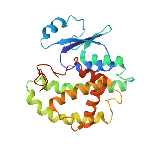
    Glutathione S-transferases (GSTs) represent the major class of detoxifying enzymes from parasitic helminths. As a result, they are candidates for chemotherapeutic and vaccine design. Indeed, GSTs from Fasciola hepatica have been found to be effective for vaccinating sheep and cattle against fasciolosis. This helminth contains at least seven GST isoforms, of which four have been cloned. The cloned isoforms (Fh51, Fh47, Fh7 and Fh1) all belong to the mu class of GSTs, share greater than 71% sequence identity, yet display distinct substrate specificities. Crystals of Fh47 were obtained using the hanging drop vapour diffusion technique. The crystals belong to space group I4122, with one monomer in the asymmetric unit, which corresponds to a very high solvent content of approximately 75%. The physiological dimer is generated via a crystallographic 2-fold rotation. The three-dimensional structure of Fh47 was solved by molecular replacement using the Schistosoma japonicum glutathione S-transferase (Sj26) crystal structure as a search model. The structure adopts the canonical GST fold comprising two domains: an N-terminal glutathione-binding domain, consisting of a four-stranded beta-sheet and three helices whilst the C-terminal domain is entirely alpha-helical. The presence of Phe19 in Fh47 results in a 6 degrees interdomain rotation in comparison to Sj26, where the equivalent residue is a leucine. Homology models of Fh51, Fh7 and Fh1, based on the Fh47 crystal structure, reveal critical differences in the residues lining the xenobiotic binding site, particularly at residue positions 9, 106 and 204. In addition, differences amongst the isoforms in the non-substrate binding site were noted, which may explain the observed differential binding of large ligands. The major immunogenic epitopes of Fh47 were surprisingly found not to reside on the most solvent-exposed regions of the molecule.


  • Organizational Affiliation
    • The Ian Potter Foundation Protein Crystallography Laboratory, St Vincent's Institute of Medical Research, 41 Victoria Parade, Fitzroy, Victoria, 3065, Australia.

Macromolecule Content 

  • Total Structure Weight: 50.92 kDa 
  • Atom Count: 3,760 
  • Modeled Residue Count: 432 
  • Deposited Residue Count: 432 
  • Unique protein chains: 1

Macromolecules

Find similar proteins by:|  3D Structure
Entity ID: 1
MoleculeChains  Sequence LengthOrganismDetailsImage
GLUTATHIONE S-TRANSFERASE
A, B
216Fasciola hepaticaMutation(s): 0 
EC: 2.5.1.18
UniProt
Find proteins for P56598 (Fasciola hepatica)
Explore P56598 
Go to UniProtKB:  P56598
Entity Groups
Sequence Clusters30% Identity50% Identity70% Identity90% Identity95% Identity100% Identity
UniProt GroupP56598
Sequence Annotations
Expand
Reference Sequence

Small Molecules

Ligands 1 Unique
IDChains Name / Formula / InChI Key2D Diagram3D Interactions
GSH

Query on GSH


Download:Ideal Coordinates CCD File
C [auth A],
D [auth B]
Glutathione
C10 H17 N3 O6 S
RWSXRVCMGQZWBV-WDSKDSINSA-N

Biologically Interesting Molecules (External Reference) 

1 Unique
Entity ID: 2
IDChains NameType/Class2D Diagram3D Interactions
PRD_002593 (GSH)
Query on PRD_002593
C [auth A],
D [auth B]
GlutathionePeptide-like / Oxidation-reduction

Experimental Data & Validation

Experimental Data

  • Method: X-RAY DIFFRACTION
  • Resolution: 2.30 Å
  • R-Value Free:  0.234 (Depositor) 
  • R-Value Work:  0.183 (Depositor) 
  • R-Value Observed: 0.183 (Depositor) 
Space Group: P 21 21 21
Unit Cell:
Length ( Å )Angle ( ˚ )
a = 62.2α = 90
b = 80.3β = 90
c = 105.9γ = 90
Software Package:
Software NamePurpose
AMoREphasing
X-PLORrefinement
DENZOdata reduction
SCALEPACKdata scaling

Structure Validation

View Full Validation Report


Entry History 

Deposition Data

Revision History  (Full details and data files)

  • Version 1.0: 1998-10-28
    Type: Initial release
  • Version 1.1: 2008-04-27
    Changes: Version format compliance
  • Version 1.2: 2011-07-13
    Changes: Database references, Derived calculations, Non-polymer description, Version format compliance
  • Version 1.3: 2011-12-21
    Changes: Non-polymer description, Structure summary
  • Version 1.4: 2023-08-30
    Changes: Data collection, Database references, Derived calculations, Refinement description