2FD2 | pdb_00002fd2

CRYSTALLOGRAPHIC ANALYSIS OF TWO SITE-DIRECTED MUTANTS OF AZOTOBACTER VINELANDII FERREDOXIN


Experimental Data Snapshot

  • Method: X-RAY DIFFRACTION
  • Resolution: 1.90 Å
  • R-Value Work: 
    0.232 (Depositor) 

wwPDB Validation 3D Report Full Report

Validation slider image for 2FD2

This is version 1.3 of the entry. See complete history

Literature

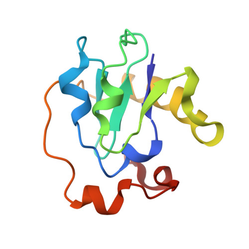
Crystallographic analysis of two site-directed mutants of Azotobacter vinelandii ferredoxin.

Soman, J.Iismaa, S.Stout, C.D.

(1991) J Biological Chem 266: 21558-21562

  • Primary Citation Related Structures: 
    2FD2

  • PubMed Abstract: 

    The crystal structure of the C24A mutant of Azotobacter vinelandii 7Fe ferredoxin (FdI) has been solved and refined at 2.0-A resolution. The structure is isomorphous to native FdI except at the site of mutation where A24 moves toward the [4Fe-4S] cluster. In spite of this inefficient packing results: three of five van der Waals contacts from the S gamma of C24 in native FdI are lost and the remaining two become longer. Consequently, the [4Fe-4S] cluster is either disordered or has a higher temperature factor (B factor) compared to the rest of the C24A FdI molecule. In addition, the entire C24A FdI structure has a higher overall B factor than native FdI. Therefore, in comparison to native FdI, the C24A mutant is isomorphous but exhibits large differences in B factor, especially at the [4Fe-4S] cluster. In contrast, the C20A FdI structure (Martin, A. G., Burgess, B. K., Stout, C. D., Cash, V. L., Dean, D. R., Jensen, G. M., and Stephens, P. J. (1990) Proc. Natl. Acad. Sci. U. S. A. 87, 598-602), which contains large structural rearrangements in the vicinity of the [4Fe-4S] cluster, exhibits essentially no change in B factor. The conformational change observed at residue 24 is similar in both C24A and C20A FdI structures. The solvent accessibility of the Fe atoms in the [3Fe-4S] and [4Fe-4S] clusters is similar in C24A, C20A, and native FdI.


  • Organizational Affiliation
    • Department of Molecular Biology, Research Institute of Scripps Clinic, La Jolla, California 92037.

Macromolecule Content 

  • Total Structure Weight: 12.67 kDa 
  • Atom Count: 885 
  • Modeled Residue Count: 106 
  • Deposited Residue Count: 106 
  • Unique protein chains: 1

Macromolecules

Find similar proteins by:|  3D Structure
Entity ID: 1
MoleculeChains  Sequence LengthOrganismDetailsImage
FERREDOXIN106Azotobacter vinelandiiMutation(s): 0 
UniProt
Find proteins for P00214 (Azotobacter vinelandii)
Explore P00214 
Go to UniProtKB:  P00214
Entity Groups
Sequence Clusters30% Identity50% Identity70% Identity90% Identity95% Identity100% Identity
UniProt GroupP00214
Sequence Annotations
Expand
Reference Sequence

Small Molecules

Ligands 2 Unique
IDChains Name / Formula / InChI Key2D Diagram3D Interactions
SF4

Query on SF4


Download:Ideal Coordinates CCD File
B [auth A]IRON/SULFUR CLUSTER
Fe4 S4
LJBDFODJNLIPKO-UHFFFAOYSA-N
F3S

Query on F3S


Download:Ideal Coordinates CCD File
C [auth A]FE3-S4 CLUSTER
Fe3 S4
FCXHZBQOKRZXKS-UHFFFAOYSA-N

Experimental Data & Validation

Experimental Data

  • Method: X-RAY DIFFRACTION
  • Resolution: 1.90 Å
  • R-Value Work:  0.232 (Depositor) 
Space Group: P 41 21 2
Unit Cell:
Length ( Å )Angle ( ˚ )
a = 55.2α = 90
b = 55.2β = 90
c = 95.2γ = 90
Software Package:
Software NamePurpose
X-PLORrefinement

Structure Validation

View Full Validation Report


Entry History 

Deposition Data

  • Released Date: 1990-10-15 
  • Deposition Author(s): Stout, C.D.

Revision History  (Full details and data files)

  • Version 1.0: 1990-10-15
    Type: Initial release
  • Version 1.1: 2008-03-24
    Changes: Version format compliance
  • Version 1.2: 2011-07-13
    Changes: Version format compliance
  • Version 1.3: 2024-02-14
    Changes: Data collection, Database references, Derived calculations, Other