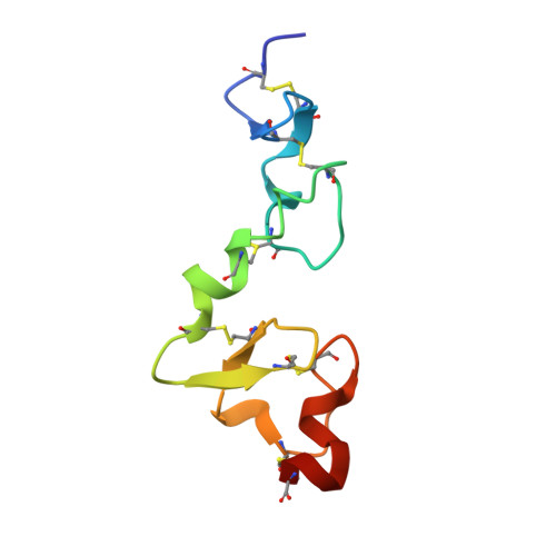
2FCW

Structure of a Complex Between the Pair of the LDL Receptor Ligand-Binding Modules 3-4 and the Receptor Associated Protein (RAP).


Experimental Data Snapshot

  • Method: X-RAY DIFFRACTION
  • Resolution: 1.26 Å
  • R-Value Free: 0.200 
  • R-Value Work: 0.152 
  • R-Value Observed: 0.135 

Starting Model: other
View more details

wwPDB Validation   3D Report Full Report


This is version 1.5 of the entry. See complete history


Literature

Structure of an LDLR-RAP Complex Reveals a General Mode for Ligand Recognition by Lipoprotein Receptors

Fisher, C.Beglova, N.Blacklow, S.C.

(2006) Mol Cell 22: 277-283

  • DOI: https://doi.org/10.1016/j.molcel.2006.02.021
  • Primary Citation of Related Structures:  
    2FCW

  • PubMed Abstract: 

    Proteins of the low-density lipoprotein receptor (LDLR) family are remarkable in their ability to bind an extremely diverse range of protein and lipoprotein ligands, yet the basis for ligand recognition is poorly understood. Here, we report the 1.26 A X-ray structure of a complex between a two-module region of the ligand binding domain of the LDLR and the third domain of RAP, an escort protein for LDLR family members. The RAP domain forms a three-helix bundle with two docking sites, one for each LDLR module. The mode of recognition at each site is virtually identical: three conserved, calcium-coordinating acidic residues from each LDLR module encircle a lysine side chain protruding from the second helix of RAP. This metal-dependent mode of electrostatic recognition, together with avidity effects resulting from the use of multiple sites, represents a general binding strategy likely to apply in the binding of other basic ligands to LDLR family proteins.


  • Organizational Affiliation

    Department of Pathology, Brigham and Women's Hospital, Harvard Medical School, 77 Avenue Louis Pasteur, Boston, Massachusetts 02115, USA.


Macromolecules
Find similar proteins by:  (by identity cutoff)  |  3D Structure
Entity ID: 1
MoleculeChains Sequence LengthOrganismDetailsImage
Alpha-2-macroglobulin receptor-associated protein109Homo sapiensMutation(s): 1 
UniProt & NIH Common Fund Data Resources
Find proteins for P30533 (Homo sapiens)
Explore P30533 
Go to UniProtKB:  P30533
PHAROS:  P30533
GTEx:  ENSG00000163956 
Entity Groups  
Sequence Clusters30% Identity50% Identity70% Identity90% Identity95% Identity100% Identity
UniProt GroupP30533
Sequence Annotations
Expand
  • Reference Sequence
Find similar proteins by:  (by identity cutoff)  |  3D Structure
Entity ID: 2
MoleculeChains Sequence LengthOrganismDetailsImage
Low-density lipoprotein receptor80Homo sapiensMutation(s): 0 
Gene Names: LDLR
UniProt & NIH Common Fund Data Resources
Find proteins for P01130 (Homo sapiens)
Explore P01130 
Go to UniProtKB:  P01130
PHAROS:  P01130
GTEx:  ENSG00000130164 
Entity Groups  
Sequence Clusters30% Identity50% Identity70% Identity90% Identity95% Identity100% Identity
UniProt GroupP01130
Sequence Annotations
Expand
  • Reference Sequence
Small Molecules
Ligands 3 Unique
IDChains Name / Formula / InChI Key2D Diagram3D Interactions
MPD
Query on MPD

Download Ideal Coordinates CCD File 
F [auth A],
G [auth A],
K [auth B],
L [auth B],
M [auth B]
(4S)-2-METHYL-2,4-PENTANEDIOL
C6 H14 O2
SVTBMSDMJJWYQN-YFKPBYRVSA-N
CA
Query on CA

Download Ideal Coordinates CCD File 
H [auth B],
I [auth B]
CALCIUM ION
Ca
BHPQYMZQTOCNFJ-UHFFFAOYSA-N
NA
Query on NA

Download Ideal Coordinates CCD File 
C [auth A],
D [auth A],
E [auth A],
J [auth B]
SODIUM ION
Na
FKNQFGJONOIPTF-UHFFFAOYSA-N
Experimental Data & Validation

Experimental Data

  • Method: X-RAY DIFFRACTION
  • Resolution: 1.26 Å
  • R-Value Free: 0.200 
  • R-Value Work: 0.152 
  • R-Value Observed: 0.135 
  • Space Group: C 1 2 1
Unit Cell:
Length ( Å )Angle ( ˚ )
a = 118.523α = 90
b = 30.019β = 96.57
c = 58.143γ = 90
Software Package:
Software NamePurpose
SHELXmodel building
SHELXL-97refinement
HKL-2000data reduction
SCALEPACKdata scaling
SOLVEphasing

Structure Validation

View Full Validation Report


Entry History 

Deposition Data

Revision History  (Full details and data files)

  • Version 1.0: 2006-05-16
    Type: Initial release
  • Version 1.1: 2008-05-01
    Changes: Version format compliance
  • Version 1.2: 2011-07-13
    Changes: Version format compliance
  • Version 1.3: 2021-10-20
    Changes: Database references, Derived calculations
  • Version 1.4: 2024-04-03
    Changes: Data collection, Refinement description
  • Version 1.5: 2024-11-06
    Changes: Structure summary