2FB3 | pdb_00002fb3

Structure of MoaA in complex with 5'-GTP


Experimental Data Snapshot

  • Method: X-RAY DIFFRACTION
  • Resolution: 2.35 Å
  • R-Value Free: 
    0.271 (Depositor), 0.266 (DCC) 
  • R-Value Work: 
    0.220 (Depositor), 0.215 (DCC) 
  • R-Value Observed: 
    0.229 (Depositor) 

wwPDB Validation 3D Report Full Report

Validation slider image for 2FB3

Ligand Structure Quality Assessment 


This is version 1.3 of the entry. See complete history

Literature

Binding of 5'-GTP to the C-terminal FeS cluster of the radical S-adenosylmethionine enzyme MoaA provides insights into its mechanism

Haenzelmann, P.Schindelin, H.

(2006) Proc Natl Acad Sci U S A 103: 6829-6834

  • DOI: https://doi.org/10.1073/pnas.0510711103
  • Primary Citation Related Structures: 
    2FB2, 2FB3

  • PubMed Abstract: 

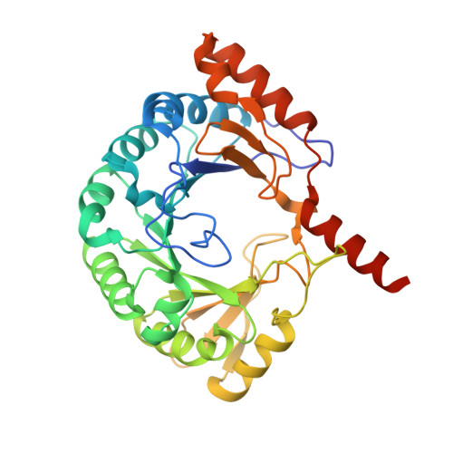
    The first step in molybdenum cofactor biosynthesis, the conversion of 5'-GTP to precursor Z, an oxygen-sensitive tetrahydropyranopterin is catalyzed by the S-adenosylmethionine (SAM)-dependent enzyme MoaA and the accessory protein MoaC. This reaction involves the radical-initiated intramolecular rearrangement of the guanine C8 atom. MoaA harbors an N-terminal [4Fe-4S] cluster, which is involved in the reductive cleavage of SAM and generates a 5'-deoxyadenosyl radical (5'-dA*), and a C-terminal [4Fe-4S] cluster presumably involved in substrate binding and/or activation. Biochemical studies identified residues involved in 5'-GTP binding and the determinants of nucleotide specificity. The crystal structure of MoaA in complex with 5'-GTP confirms the biochemical data and provides valuable insights into the subsequent radical reaction. MoaA binds 5'-GTP with high affinity and interacts through its C-terminal [4Fe-4S] cluster with the guanine N1 and N2 atoms, in a yet uncharacterized binding mode. The tightly anchored triphosphate moiety prevents the escape of radical intermediates. This structure also visualizes the L-Met and 5'-dA cleavage products of SAM. Rotation of the 5'-dA ribose and/or conformational changes of the guanosine are proposed to bring the 5'-deoxyadenosyl radical into close proximity of either the ribose C2' and C3' or the guanine C8 carbon atoms leading to hydrogen abstraction.


  • Organizational Affiliation
    • Department of Biochemistry and Center for Structural Biology, Stony Brook University, Stony Brook, NY 11794-5215, USA. petra@csb.sunysb.edu

Macromolecule Content 

  • Total Structure Weight: 81.11 kDa 
  • Atom Count: 5,518 
  • Modeled Residue Count: 653 
  • Deposited Residue Count: 680 
  • Unique protein chains: 1

Macromolecules

Find similar proteins by:|  3D Structure
Entity ID: 1
MoleculeChains  Sequence LengthOrganismDetailsImage
Molybdenum cofactor biosynthesis protein A
A, B
340Staphylococcus aureusMutation(s): 0 
Gene Names: MoaA
EC: 4.1.99.22
UniProt
Find proteins for P69848 (Staphylococcus aureus (strain NCTC 8325 / PS 47))
Explore P69848 
Go to UniProtKB:  P69848
Entity Groups
Sequence Clusters30% Identity50% Identity70% Identity90% Identity95% Identity100% Identity
UniProt GroupP69848
Sequence Annotations
Expand
Reference Sequence

Small Molecules

Ligands 6 Unique
IDChains Name / Formula / InChI Key2D Diagram3D Interactions
GTP

Query on GTP


Download:Ideal Coordinates CCD File
G [auth A]GUANOSINE-5'-TRIPHOSPHATE
C10 H16 N5 O14 P3
XKMLYUALXHKNFT-UUOKFMHZSA-N
SF4

Query on SF4


Download:Ideal Coordinates CCD File
E [auth A],
F [auth A],
K [auth B],
L [auth B]
IRON/SULFUR CLUSTER
Fe4 S4
LJBDFODJNLIPKO-UHFFFAOYSA-N
5AD

Query on 5AD


Download:Ideal Coordinates CCD File
H [auth A]5'-DEOXYADENOSINE
C10 H13 N5 O3
XGYIMTFOTBMPFP-KQYNXXCUSA-N
POP

Query on POP


Download:Ideal Coordinates CCD File
M [auth B]PYROPHOSPHATE 2-
H2 O7 P2
XPPKVPWEQAFLFU-UHFFFAOYSA-L
MET

Query on MET


Download:Ideal Coordinates CCD File
D [auth A],
J [auth B]
METHIONINE
C5 H11 N O2 S
FFEARJCKVFRZRR-BYPYZUCNSA-N
SO4

Query on SO4


Download:Ideal Coordinates CCD File
C [auth A],
I [auth B]
SULFATE ION
O4 S
QAOWNCQODCNURD-UHFFFAOYSA-L

Experimental Data & Validation

Experimental Data

  • Method: X-RAY DIFFRACTION
  • Resolution: 2.35 Å
  • R-Value Free:  0.271 (Depositor), 0.266 (DCC) 
  • R-Value Work:  0.220 (Depositor), 0.215 (DCC) 
  • R-Value Observed: 0.229 (Depositor) 
Space Group: P 21 21 21
Unit Cell:
Length ( Å )Angle ( ˚ )
a = 48.015α = 90
b = 103.449β = 90
c = 191.217γ = 90
Software Package:
Software NamePurpose
CBASSdata collection
SCALEPACKdata scaling
FFTmodel building
REFMACrefinement
FFTphasing

Structure Validation

View Full Validation Report


Ligand Structure Quality Assessment 


Entry History 

Deposition Data

Revision History  (Full details and data files)

  • Version 1.0: 2006-05-09
    Type: Initial release
  • Version 1.1: 2008-05-01
    Changes: Version format compliance
  • Version 1.2: 2011-07-13
    Changes: Advisory, Version format compliance
  • Version 1.3: 2024-02-14
    Changes: Data collection, Database references, Derived calculations, Refinement description