2F9D

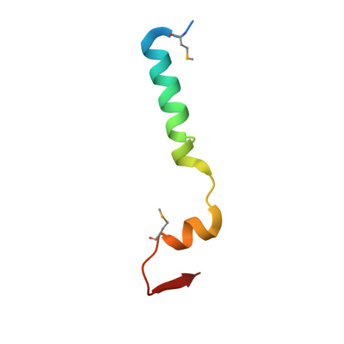
2.5 angstrom resolution structure of the spliceosomal protein p14 bound to region of SF3b155


Experimental Data Snapshot

  • Method: X-RAY DIFFRACTION
  • Resolution: 2.50 Å
  • R-Value Free: 0.276 
  • R-Value Work: 0.221 
  • R-Value Observed: 0.224 

wwPDB Validation   3D Report Full Report


This is version 1.4 of the entry. See complete history


Literature

Crystal structure of a core spliceosomal protein interface

Schellenberg, M.J.Edwards, R.A.Ritchie, D.B.Kent, O.A.Golas, M.M.Stark, H.Glover, J.N.M.Macmillan, A.M.

(2006) Proc Natl Acad Sci U S A 103: 1266-1271

  • DOI: https://doi.org/10.1073/pnas.0508048103
  • Primary Citation of Related Structures:  
    2F9D, 2F9J

  • PubMed Abstract: 

    The precise excision of introns from precursor mRNAs (pre-mRNAs) in eukaryotes is accomplished by the spliceosome, a complex assembly containing five small nuclear ribonucleoprotein (snRNP) particles. Human p14, a component of the spliceosomal U2 and U11/U12 snRNPs, has been shown to associate directly with the pre-mRNA branch adenosine early in spliceosome assembly and within the fully assembled spliceosome. Here we report the 2.5-A crystal structure of a complex containing p14 and a peptide derived from the p14-associated U2 snRNP component SF3b155. p14 contains an RNA recognition motif (RRM), the surface of which is largely occluded by a C-terminal alpha-helix and a portion of the SF3b155 peptide. An analysis of RNA.protein crosslinking to wild-type and mutant p14 shows that the branch adenosine directly interacts with a conserved aromatic within a pocket on the surface of the complex. This result, combined with a comparison of the structure with known RRMs and pseudoRRMs as well as model-building by using the electron cryomicroscopy structure of a spliceosomal U11/U12 di-snRNP, suggests that p14.SF3b155 presents a noncanonical surface for RNA recognition at the heart of the mammalian spliceosome.


  • Organizational Affiliation

    Department of Biochemistry, University of Alberta, Edmonton, Alberta, Canada T6G 2H7.


Macromolecules
Find similar proteins by:  (by identity cutoff)  |  3D Structure
Entity ID: 1
MoleculeChains Sequence LengthOrganismDetailsImage
Pre-mRNA branch site protein p14
A, B
125Homo sapiensMutation(s): 0 
Gene Names: SF3B14
UniProt & NIH Common Fund Data Resources
Find proteins for Q9Y3B4 (Homo sapiens)
Go to UniProtKB:  Q9Y3B4
PHAROS:  Q9Y3B4
GTEx:  ENSG00000115128 
Sequence Annotations
Expand
  • Reference Sequence
Find similar proteins by:  (by identity cutoff)  |  3D Structure
Entity ID: 2
MoleculeChains Sequence LengthOrganismDetailsImage
Splicing factor 3B subunit 1C [auth P],
D [auth Q]
43Homo sapiensMutation(s): 3 
Gene Names: SF3B1SAP155
UniProt & NIH Common Fund Data Resources
Find proteins for O75533 (Homo sapiens)
Go to UniProtKB:  O75533
PHAROS:  O75533
GTEx:  ENSG00000115524 
Sequence Annotations
Expand
  • Reference Sequence
Small Molecules
Modified Residues  1 Unique
IDChains TypeFormula2D DiagramParent
MSE
Query on MSE
C [auth P],
D [auth Q]
L-PEPTIDE LINKINGC5 H11 N O2 SeMET
Experimental Data & Validation

Experimental Data

  • Method: X-RAY DIFFRACTION
  • Resolution: 2.50 Å
  • R-Value Free: 0.276 
  • R-Value Work: 0.221 
  • R-Value Observed: 0.224 
  • Space Group: C 2 2 21
Unit Cell:
Length ( Å )Angle ( ˚ )
a = 101.87α = 90
b = 115.227β = 90
c = 82.536γ = 90
Software Package:
Software NamePurpose
DENZOdata reduction
SCALEPACKdata scaling
SOLVEphasing
RESOLVEphasing
REFMACrefinement
PDB_EXTRACTdata extraction
MOSFLMdata reduction

Structure Validation

View Full Validation Report


Entry History 

Deposition Data

Revision History  (Full details and data files)

  • Version 1.0: 2006-01-24
    Type: Initial release
  • Version 1.1: 2008-05-01
    Changes: Version format compliance
  • Version 1.2: 2011-07-13
    Changes: Advisory, Refinement description, Version format compliance
  • Version 1.3: 2017-10-18
    Changes: Refinement description
  • Version 1.4: 2024-10-30
    Changes: Data collection, Database references, Derived calculations, Refinement description, Structure summary