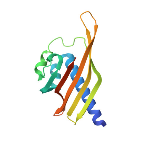
2F86

The Association Domain of C. elegans CaMKII


Experimental Data Snapshot

  • Method: X-RAY DIFFRACTION
  • Resolution: 2.64 Å
  • R-Value Free: 0.301 
  • R-Value Work: 0.248 
  • R-Value Observed: 0.248 

Starting Model: experimental
View more details

wwPDB Validation   3D Report Full Report


This is version 1.5 of the entry. See complete history


Literature

Oligomerization states of the association domain and the holoenyzme of Ca2+/CaM kinase II.

Rosenberg, O.S.Deindl, S.Comolli, L.R.Hoelz, A.Downing, K.H.Nairn, A.C.Kuriyan, J.

(2006) FEBS J 273: 682-694

  • DOI: https://doi.org/10.1111/j.1742-4658.2005.05088.x
  • Primary Citation of Related Structures:  
    2F86

  • PubMed Abstract: 

    Ca2+/calmodulin activated protein kinase II (CaMKII) is an oligomeric protein kinase with a unique holoenyzme architecture. The subunits of CaMKII are bound together into the holoenzyme by the association domain, a C-terminal region of approximately 140 residues in the CaMKII polypeptide. Single particle analyses of electron micrographs have suggested previously that the holoenyzme forms a dodecamer that contains two stacked 6-fold symmetric rings. In contrast, a recent crystal structure of the isolated association domain of mouse CaMKIIalpha has revealed a tetradecameric assembly with two stacked 7-fold symmetric rings. In this study, we have determined the crystal structure of the Caenorhabditis elegans CaMKII association domain and it too forms a tetradecamer. We also show by electron microscopy that in its fully assembled form the CaMKII holoenzyme is a dodecamer but without the kinase domains, either from expression of the isolated association domain in bacteria or following their removal by proteolysis, the association domains form a tetradecamer. We speculate that the holoenzyme is held in its 6-fold symmetric state by the interactions of the N-terminal approximately 1-335 residues and that the removal of this region allows the association domain to convert into a more stable 7-fold symmetric form.


  • Organizational Affiliation

    Department of Molecular and Cell Biology, University of California, Berkeley, CA 94720-3202, USA.


Macromolecules
Find similar proteins by:  (by identity cutoff)  |  3D Structure
Entity ID: 1
MoleculeChains Sequence LengthOrganismDetailsImage
Hypothetical protein K11E8.1d143Caenorhabditis elegansMutation(s): 0 
Gene Names: unc-43K11E8.1
EC: 2.7.11.17
UniProt
Find proteins for O62305 (Caenorhabditis elegans)
Explore O62305 
Go to UniProtKB:  O62305
Entity Groups  
Sequence Clusters30% Identity50% Identity70% Identity90% Identity95% Identity100% Identity
UniProt GroupO62305
Sequence Annotations
Expand
  • Reference Sequence
Experimental Data & Validation

Experimental Data

  • Method: X-RAY DIFFRACTION
  • Resolution: 2.64 Å
  • R-Value Free: 0.301 
  • R-Value Work: 0.248 
  • R-Value Observed: 0.248 
  • Space Group: C 2 2 21
Unit Cell:
Length ( Å )Angle ( ˚ )
a = 70.87α = 90
b = 186.934β = 90
c = 182.934γ = 90
Software Package:
Software NamePurpose
CNSrefinement
MADNESSdata reduction
SCALEPACKdata scaling
PHASERphasing

Structure Validation

View Full Validation Report


Entry History 

Deposition Data

Revision History  (Full details and data files)

  • Version 1.0: 2006-02-07
    Type: Initial release
  • Version 1.1: 2008-05-01
    Changes: Version format compliance
  • Version 1.2: 2011-07-13
    Changes: Derived calculations, Version format compliance
  • Version 1.3: 2017-06-28
    Changes: Database references, Source and taxonomy, Structure summary
  • Version 1.4: 2017-10-18
    Changes: Refinement description
  • Version 1.5: 2023-08-30
    Changes: Data collection, Database references, Refinement description