2F55 | pdb_00002f55

Two hepatitis c virus ns3 helicase domains complexed with the same strand of dna


Experimental Data Snapshot

  • Method: X-RAY DIFFRACTION
  • Resolution: 3.30 Å
  • R-Value Free: 
    0.273 (Depositor) 
  • R-Value Work: 
    0.247 (Depositor) 
  • R-Value Observed: 
    0.247 (Depositor) 

Starting Model: experimental
View more details

wwPDB Validation 3D Report Full Report

Validation slider image for 2F55

This is version 1.4 of the entry. See complete history

Literature

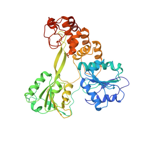
Structural and biological identification of residues on the surface of NS3 helicase required for optimal replication of the hepatitis C virus

Mackintosh, S.G.Lu, J.Z.Jordan, J.B.Harrison, M.K.Sikora, B.Sharma, S.D.Cameron, C.E.Raney, K.D.Sakon, J.

(2006) J Biological Chem 281: 3528-3535

  • DOI: https://doi.org/10.1074/jbc.M512100200
  • Primary Citation Related Structures: 
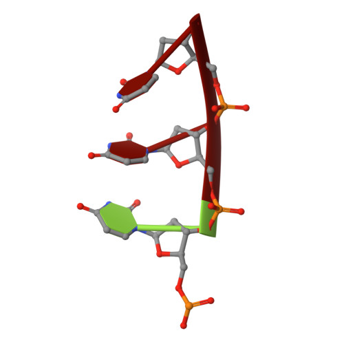
    2F55

  • PubMed Abstract: 

    The hepatitis C virus (HCV) nonstructural protein 3 (NS3) is a multifunctional enzyme with serine protease and DEXH/D-box helicase domains. A crystal structure of the NS3 helicase domain (NS3h) was generated in the presence of a single-stranded oligonucleotide long enough to accommodate binding of two molecules of enzyme. Several amino acid residues at the interface of the two NS3h molecules were identified that appear to mediate a protein-protein interaction between domains 2 and 3 of adjacent molecules. Mutations were introduced into domain 3 to disrupt the putative interface and subsequently examined using an HCV subgenomic replicon, resulting in significant reduction in replication capacity. The mutations in domain 3 were then examined using recombinant NS3h in biochemical assays. The mutant enzyme showed RNA binding and RNA-stimulated ATPase activity that mirrored wild type NS3h. In DNA unwinding assays under single turnover conditions, the mutant NS3h exhibited a similar unwinding rate and only approximately 2-fold lower processivity than wild type NS3h. Overall biochemical activities of the mutant NS3h were similar to the wild type enzyme, which was not reflective of the large reduction in HCV replicative capacity observed in the biological experiment. Hence, the biological results suggest that the known biochemical properties associated with the helicase activity of NS3h do not reveal all of the likely biological roles of NS3 during HCV replication. Domain 3 of NS3 is implicated in protein-protein interactions that are necessary for HCV replication.


  • Organizational Affiliation
    • Department of Biochemistry and Molecular Biology, University of Arkansas for Medical Sciences, Little Rock, Arkansas 72205, USA.

Macromolecule Content 

  • Total Structure Weight: 144.46 kDa 
  • Atom Count: 10,129 
  • Modeled Residue Count: 1,312 
  • Deposited Residue Count: 1,321 
  • Unique protein chains: 1
  • Unique nucleic acid chains: 2

Macromolecules


Find similar proteins by:|  3D Structure
Entity ID: 3
MoleculeChains  Sequence LengthOrganismDetailsImage
polyproteinC [auth A],
D [auth B],
E [auth C]
435Orthohepacivirus hominisMutation(s): 0 
EC: 3.6.1
UniProt
Find proteins for O92972 (Hepatitis C virus genotype 1b (strain HC-J4))
Explore O92972 
Go to UniProtKB:  O92972
Entity Groups
Sequence Clusters30% Identity50% Identity70% Identity90% Identity95% Identity100% Identity
UniProt GroupO92972
Sequence Annotations
Expand
Reference Sequence
Find similar nucleic acids by:  Sequence
Entity ID: 1
MoleculeChains LengthOrganismImage
5'-D(P*(DU)P*(DU)P*(DU)P*(DU)P*(DU)P*(DU)P*(DU)P*(DU)P*(DU)P*(DU)P*(DU)P*(DU)P*(DU))-3'A [auth D]13N/A
Sequence Annotations
Expand
Reference Sequence
Find similar nucleic acids by:  Sequence
Entity ID: 2
MoleculeChains LengthOrganismImage
5'-D(P*(DU)P*(DU)P*(DU))-3'B [auth E]3N/A
Sequence Annotations
Expand
Reference Sequence

Small Molecules

Experimental Data & Validation

Experimental Data

  • Method: X-RAY DIFFRACTION
  • Resolution: 3.30 Å
  • R-Value Free:  0.273 (Depositor) 
  • R-Value Work:  0.247 (Depositor) 
  • R-Value Observed: 0.247 (Depositor) 
Space Group: P 21 21 21
Unit Cell:
Length ( Å )Angle ( ˚ )
a = 108.2α = 90
b = 109.8β = 90
c = 183.4γ = 90
Software Package:
Software NamePurpose
SCALEPACKdata scaling
AMoREphasing
CNSrefinement

Structure Validation

View Full Validation Report


Entry History 

Deposition Data

Revision History  (Full details and data files)

  • Version 1.0: 2005-12-06
    Type: Initial release
  • Version 1.1: 2008-05-01
    Changes: Version format compliance
  • Version 1.2: 2011-07-13
    Changes: Version format compliance
  • Version 1.3: 2017-10-18
    Changes: Refinement description
  • Version 1.4: 2023-08-23
    Changes: Data collection, Database references, Derived calculations, Refinement description