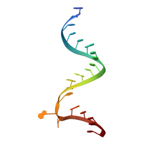
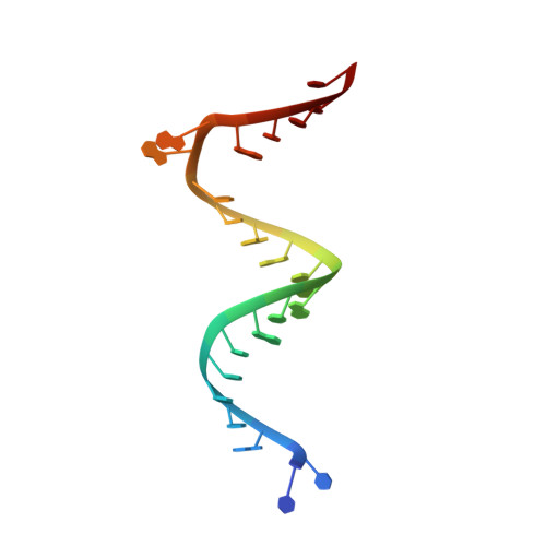
2F4T

Asite RNA + designer antibiotic


Experimental Data Snapshot

  • Method: X-RAY DIFFRACTION
  • Resolution: 3.00 Å
  • R-Value Free: 0.267 
  • R-Value Work: 0.211 
  • R-Value Observed: 0.267 

wwPDB Validation   3D Report Full Report


This is version 1.4 of the entry. See complete history


Literature

Interactions of designer antibiotics and the bacterial ribosomal aminoacyl-tRNA site

Murray, J.B.Meroueh, S.O.Russell, R.J.Lentzen, G.Haddad, J.Mobashery, S.

(2006) Chem Biol 13: 129-138

  • DOI: https://doi.org/10.1016/j.chembiol.2005.11.004
  • Primary Citation of Related Structures:  
    2F4S, 2F4T, 2F4U, 2F4V

  • PubMed Abstract: 

    The X-ray crystal structures for the complexes of three designer antibiotics, compounds 1, 2, and 3, bound to two models for the ribosomal aminoacyl-tRNA site (A site) at 2.5-3.0 Angstroms resolution and that of neamine at 2.8 Angstroms resolution are described. Furthermore, the complex of antibiotic 1 bound to the A site in the entire 30S ribosomal subunit of Thermus thermophilus is reported at 3.8 Angstroms resolution. Molecular dynamics simulations revealed that the designer compounds provide additional stability to bases A1492 and A1493 in their extrahelical forms. Snapshots from the simulations were used for free energy calculations, which revealed that van der Waals and hydrophobic effects were the driving forces behind the binding of designer antibiotic 3 when compared to the parental neamine.


  • Organizational Affiliation

    Vernalis (R&D), Ltd., Granta Park, Cambridge, UK. j.murray@vernalis.com


Macromolecules

Find similar nucleic acids by:  Sequence   |   3D Structure  

Entity ID: 1
MoleculeChains LengthOrganismImage
5'-R(*GP*CP*GP*UP*CP*AP*CP*AP*CP*CP*GP*GP*UP*GP*AP*AP*GP*UP*CP*GP*C)-3'21N/A
Sequence Annotations
Expand
  • Reference Sequence

Find similar nucleic acids by:  Sequence   |   3D Structure  

Entity ID: 2
MoleculeChains LengthOrganismImage
5'-R(*UP*UP*GP*CP*GP*UP*CP*AP*CP*AP*CP*CP*GP*GP*UP*GP*AP*AP*GP*UP*CP*GP*C)-3'23N/A
Sequence Annotations
Expand
  • Reference Sequence
Small Molecules
Ligands 1 Unique
IDChains Name / Formula / InChI Key2D Diagram3D Interactions
AB9
Query on AB9

Download Ideal Coordinates CCD File 
C [auth B](2R)-4-AMINO-N-{(1R,2S,3R,4R,5S)-5-AMINO-2-{2-[(2-AMINOETHYL)AMINO]ETHOXY}-4-[(2,6-DIAMINO-2,6-DIDEOXY-ALPHA-D-GLUCOPYRANOSYL)OXY]-3-HYDROXYCYCLOHEXYL}-2-HYDROXYBUTANAMIDE
C20 H43 N7 O8
OGRDTONULPXNRP-HSDDYVLRSA-N
Binding Affinity Annotations 
IDSourceBinding Affinity
AB9 PDBBind:  2F4T Kd: 2200 (nM) from 1 assay(s)
Experimental Data & Validation

Experimental Data

  • Method: X-RAY DIFFRACTION
  • Resolution: 3.00 Å
  • R-Value Free: 0.267 
  • R-Value Work: 0.211 
  • R-Value Observed: 0.267 
  • Space Group: P 1 21 1
Unit Cell:
Length ( Å )Angle ( ˚ )
a = 45.976α = 90
b = 32.926β = 95.62
c = 46.414γ = 90
Software Package:
Software NamePurpose
DENZOdata reduction
SCALEPACKdata scaling
CNSrefinement
PDB_EXTRACTdata extraction

Structure Validation

View Full Validation Report


Entry History 

Deposition Data

Revision History  (Full details and data files)

  • Version 1.0: 2006-05-02
    Type: Initial release
  • Version 1.1: 2008-05-01
    Changes: Version format compliance
  • Version 1.2: 2011-07-13
    Changes: Version format compliance
  • Version 1.3: 2017-10-18
    Changes: Advisory, Refinement description
  • Version 1.4: 2024-02-14
    Changes: Advisory, Data collection, Database references, Derived calculations