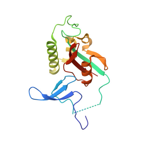
Structure-function-folding relationship in a WW domain.
Jager, M., Zhang, Y., Bieschke, J., Nguyen, H., Dendle, M., Bowman, M.E., Noel, J.P., Gruebele, M., Kelly, J.W.(2006) Proc Natl Acad Sci U S A 103: 10648-10653
- PubMed: 16807295
- DOI: https://doi.org/10.1073/pnas.0600511103
- Primary Citation of Related Structures:
1ZCN, 2F21 - PubMed Abstract:
Protein folding barriers result from a combination of factors including unavoidable energetic frustration from nonnative interactions, natural variation and selection of the amino acid sequence for function, and/or selection pressure against aggregation. The rate-limiting step for human Pin1 WW domain folding is the formation of the loop 1 substructure. The native conformation of this six-residue loop positions side chains that are important for mediating protein-protein interactions through the binding of Pro-rich sequences. Replacement of the wild-type loop 1 primary structure by shorter sequences with a high propensity to fold into a type-I' beta-turn conformation or the statistically preferred type-I G1 bulge conformation accelerates WW domain folding by almost an order of magnitude and increases thermodynamic stability. However, loop engineering to optimize folding energetics has a significant downside: it effectively eliminates WW domain function according to ligand-binding studies. The energetic contribution of loop 1 to ligand binding appears to have evolved at the expense of fast folding and additional protein stability. Thus, the two-state barrier exhibited by the wild-type human Pin1 WW domain principally results from functional requirements, rather than from physical constraints inherent to even the most efficient loop formation process.
- Department of Chemistry and The Skaggs Institute for Chemical Biology, The Scripps Research Institute, 10550 North Torrey Pines Road, BCC265, La Jolla, CA 92037, USA.
Organizational Affiliation:


