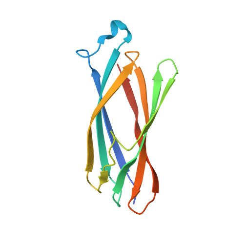
Solution structure of ApaG from Xanthomonas axonopodis pv. citri reveals a fibronectin-3 fold.
Cicero, D.O., Contessa, G.M., Pertinhez, T.A., Gallo, M., Katsuyama, A.M., Paci, M., Farah, C.S., Spisni, A.(2007) Proteins 67: 490-500
- PubMed: 17256769 Search on PubMed
- DOI: https://doi.org/10.1002/prot.21277
- Primary Citation Related Structures:
2F1E - PubMed Abstract:
ApaG proteins are found in a wide variety of bacterial genomes but their function is as yet unknown. Some eukaryotic proteins involved in protein-protein interactions, such as the human polymerase delta-interacting protein (PDIP38) and the F Box A (FBA) proteins, contain ApaG homology domains. We have used NMR to determine the solution structure of ApaG protein from the plant pathogen Xanthomonas axonopodis pv. citri (ApaG(Xac)) with the aim to shed some light on its molecular function. ApaG(Xac) is characterized by seven antiparallel beta strands forming two beta sheets, one containing three strands (ABE) and the other four strands (GFCC'). Relaxation measurements indicate that the protein has a quite rigid structure. In spite of the presence of a putative GXGXXG pyrophosphate binding motif ApaG(Xac) does not bind ATP or GTP, in vitro. On the other hand, ApaG(Xac) adopts a fibronectin type III (Fn3) fold, which is consistent with the hypothesis that it is involved in mediating protein-protein interactions. The fact that the proteins of ApaG family do not display significant sequence similarity with the Fn3 domains found in other eukaryotic or bacterial proteins suggests that Fn3 domain may have arisen earlier in evolution than previously estimated.
- Department of Chemical Science and Technology, University of Rome, Tor Vergata, Italy.
Organizational Affiliation: