2EWN

Ecoli Biotin Repressor with co-repressor analog


Experimental Data Snapshot

  • Method: X-RAY DIFFRACTION
  • Resolution: 2.80 Å
  • R-Value Free: 0.264 
  • R-Value Work: 0.213 
  • R-Value Observed: 0.214 

wwPDB Validation   3D Report Full Report


Ligand Structure Quality Assessment 


This is version 1.3 of the entry. See complete history


Literature

Co-repressor Induced Order and Biotin Repressor Dimerization: A Case for Divergent Followed by Convergent Evolution.

Wood, Z.A.Weaver, L.H.Brown, P.H.Beckett, D.Matthews, B.W.

(2006) J Mol Biol 357: 509-523

  • DOI: https://doi.org/10.1016/j.jmb.2005.12.066
  • Primary Citation of Related Structures:  
    2EWN

  • PubMed Abstract: 

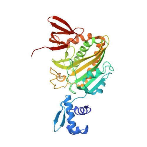
    BirA catalyzes the adenylation and subsequent covalent attachment of biotin to the biotin carboxyl carrier protein (BCCP). In the absence of apo-BCCP, biotin-5'-AMP acts as a co-repressor that induces BirA dimerization and binding to the bio operator to repress biotin biosynthesis. The crystal structures of apo-BirA, and BirA in complex with biotin have been reported. We here describe the 2.8A resolution crystal structure of BirA in complex with the co-repressor analog biotinol-5'-AMP. It was previously shown that the structure of apo-BirA is monomeric and that binding of biotin weakly induces a dimeric structure in which three disordered surface loops become organized to form the dimer interface. The structure of the co-repressor complex is also a dimer, clearly related to the BirA.biotin structure, but with several significant conformational changes. A hitherto disordered "adenylate binding loop" forms a well-defined structure covering the co-repressor. The co-repressor buttresses the dimer interface, resulting in improved packing and a 12 degrees change in the hinge-bending angle along the dimer interface relative to the BirA.biotin structure. This helps explain why the binding of the co-repressor is necessary to optimize the binding of BirA to the bioO operator. The structure reveals an unexpected use of the nucleotide-binding motif GXGXXG in binding adenylate and controlling the repressor function. Finally, based on structural analysis we propose that the class of adenylating enzymes represented by BirA, lipoate protein ligase and class II tRNA synthetases diverged early and were selected based on their ability to sequester co-factors or amino acid residues, and adenylation activity arose independently through functional convergence.


  • Organizational Affiliation

    Institute of Molecular Biology, University of Oregon, Eugene, OR 97403-1229, USA.


Macromolecules
Find similar proteins by:  (by identity cutoff)  |  3D Structure
Entity ID: 1
MoleculeChains Sequence LengthOrganismDetailsImage
BirA bifunctional protein
A, B
321Escherichia coliMutation(s): 0 
Gene Names: birAbioRdhbB
EC: 6.3.4.15
UniProt
Find proteins for P06709 (Escherichia coli (strain K12))
Explore P06709 
Go to UniProtKB:  P06709
Entity Groups  
Sequence Clusters30% Identity50% Identity70% Identity90% Identity95% Identity100% Identity
UniProt GroupP06709
Sequence Annotations
Expand
  • Reference Sequence
Small Molecules
Ligands 1 Unique
IDChains Name / Formula / InChI Key2D Diagram3D Interactions
BTX
Query on BTX

Download Ideal Coordinates CCD File 
C [auth A],
D [auth B]
((2R,3S,4R,5R)-5-(6-AMINO-9H-PURIN-9-YL)-3,4-DIHYDROXY-TETRAHYDROFURAN-2-YL)METHYL 5-((3AS,4S,6AR)-2-OXO-HEXAHYDRO-1H-THIENO[3,4-D]IMIDAZOL-4-YL)PENTYL HYDROGEN PHOSPHATE
C20 H30 N7 O8 P S
KBOGUFFJCBPJEH-SQGSUPJISA-N
Binding Affinity Annotations 
IDSourceBinding Affinity
BTX BindingDB:  2EWN Ki: 87 (nM) from 1 assay(s)
Kd: 1.5 (nM) from 1 assay(s)
Experimental Data & Validation

Experimental Data

  • Method: X-RAY DIFFRACTION
  • Resolution: 2.80 Å
  • R-Value Free: 0.264 
  • R-Value Work: 0.213 
  • R-Value Observed: 0.214 
  • Space Group: P 43 21 2
Unit Cell:
Length ( Å )Angle ( ˚ )
a = 152.188α = 90
b = 152.188β = 90
c = 93.189γ = 90
Software Package:
Software NamePurpose
CNSrefinement
DENZOdata reduction
SCALEPACKdata scaling
CNSphasing

Structure Validation

View Full Validation Report


Ligand Structure Quality Assessment 


Entry History 

Deposition Data

Revision History  (Full details and data files)

  • Version 1.0: 2006-02-21
    Type: Initial release
  • Version 1.1: 2008-05-01
    Changes: Version format compliance
  • Version 1.2: 2011-07-13
    Changes: Version format compliance
  • Version 1.3: 2023-08-23
    Changes: Data collection, Database references, Derived calculations, Refinement description